Janet L. Hart and Dr. Kim L. O’Neill, Department of Microbiology
Apoptosis is the method by which cells undergo programmed cell death or “cell suicide.” The cell initiates apoptosis after there is irreparable damage or if the cell is simply not needed. Some insults that are known to induce apoptosis are irradiation, heat shock, as well as many chemotherapeutic drugs including staurosporine, paclitaxel, and dexamethasone. The precise regulation of apoptosis is critical, as disregulation can lead to many diseases or conditions including autoimmune disorders or cancer.
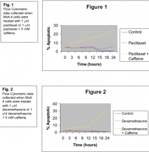
Molt-4 cells (a human T cell line) were treated simultaneously with 1 μM paclitaxel1 or 1 μM dexamethasone2 and 5 mM caffeine. Using flow cytometry, the percent of apoptotic cells was assessed by phosphatidylserine (PS) externalization (ANNEXIN V binding), which is characteristic of cells undergoing apoptosis.
The apoptotic percentage was tested at 3-hour time points for a 24-hour time course. Control cells were compared to the cells receiving the drug alone and those receiving the drug + caffeine treatment.
I chose to use the Molt-4 cell line because it is a human cell line and externalizes PS, which allows for detection of apoptosis. Paclitaxel and dexamethasone were chosen because they have different modes of action when inducing cell death. In future experiments, genotoxic drugs and insults (DNA damaging insults, such as irradiation) will be tested.
The concurrent treatment of 5 mM caffeine showed a synergistic effect with the apoptoticinducing drugs. Caffeine alone led to 10.3% of the cells undergoing apoptosis after 24 hours (data not shown). The apoptotic percentage of the control group after 24 hours was only 0.70 while the apoptotic percentage of the paclitaxel treated cells was 5.76. The paclitaxel + caffeine treated cells showed a percentage of 24.5 after the same time (see figure 1). This is more than the expected additive result of paclitaxel and caffeine.
The same trend was seen in the dexamethasone experiment. The apoptotic percentage of the control group after 24 hours was only 1.19 while the apoptotic percentage of the dexamethasone treated cells was 1.24. The dexamethasone + caffeine treated cells showed a percentage of 31.6 after the same time (see figure 2). Again, the more than additive effect is seen.
By looking at the different trends and kinetics of apoptotic induction, we can learn more about the varying pathways in the cell. The effects of caffeine on human cells have been studied, but a definite conclusion has not been reached. I would like to continue working on this experiment by testing different drugs and possibly different cell lines to see if different responses to concurrent caffeine treatment would be observed. Another interesting event during apoptotic progression is DNA fragmentation and cell cycle analysis. I think that this project is relevant to basic science and can help elucidate the role that caffeine plays in human cells that are undergoing apoptosis. This scholarship has enabled me to start this experiment, collect data and make definite observations.

