
Blake Simmons and Dr. Dixon Woodbury, Physiology and Developmental Biology
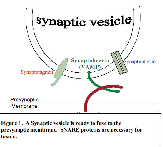
Nerve Cells communicate with each other by synaptic transmission. Synaptic transmission is the release by one nerve cell of neurotransmitters packaged in synaptic vesicles. Proteins known as SNAREs, (e.g., synaptobrevin and syntaxin) are believed to drive neurotransmitter release by inducing fusion (exocytosis) within a presynaptic neuron. Figure 1 shows a synaptic vesicle binding to the presynaptic membrane and the interaction of two SNARE proteins.i,ii My plan was to map which domains of the SNARE protein, syntaxin, are necessary for fusion.
Knowing which parts (or domains) of SNARE proteins are necessary for fusion would give us a greater fundamental understanding of nerve function. These SNARE proteins are the targets of dangerous neurotoxins produced by Clostridium tetani (tetanus) and Clostridium botulinum (botulism). Therefore, studying their function may aid in clinical treatments of tetanus and botulism.
Synaptic transmission in the nervous system is well understood except the step of neurotransmitter release. Synaptic vesicles store neurotransmitters, which are released across the presynaptic membrane by fusion. Messages are sent throughout our body with different neurotransmitters such as amino acids, amines, and peptides.
Dr. Woodbury’s lab has recently shown that native vesicles (with SNAREs in them) will fuse to membranes containing only one SNARE (syntaxin). His current work is focused on the nature of the SNARE and membrane interactions.
Preliminary data suggests that only certain parts of the syntaxin protein are necessary for fusion to occur.iii My plan was to help map which domains of syntaxin are necessary for fusion. I used truncation clones that contained shortened lengths of syntaxin to test this hypothesis.
I completed hundreds of experiments using an artificial membrane. Artificial membranes (bilayers) are membranes made out of phospholipids in a special chamber. The chamber consists of two solutions with a partition. This partition has a small hole. A pipette is used to spread a small amount of lipids over the tiny hole within the partition. The membrane (lipid film) is formed similar to a bubble of soap that forms a film in a hoop.
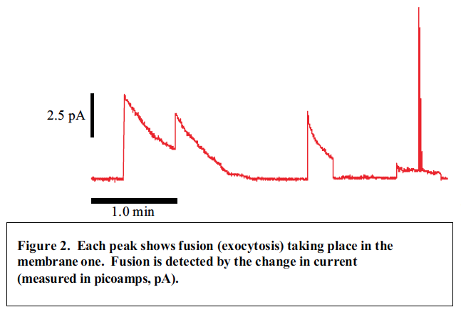
When AV’s or mSV’s fuse with a membrane a change in the electrical properties of the membrane occur. Vesicles are made with different electrical properties then the membranes. Channels or holes are placed in vesicles; when they fuse these channels are detected electrically in the membrane. Experiments I have run in Dr. Woodbury’s lab, have shown fusion occurring (Fig 2).
I was able to use two clones, Syntaxin 1A-1 and 1A-8. My experiments did not show similar results from preliminary data. This could be due to previously existing contamination or problems in growing the identical Syntaxin clones. Currently our molecular biology team, in Dr. Woodbury’s lab, is trying to purify the syntaxin and find better ways of induction to produce a greater quantity of protein. I expect that within a year, this lab will have the answer to which clones of syntaxin are more probable for fusion.
As a Neuroscience major and a future Optometrist, learning more about the function of neurons has been and is exciting. Understanding the nervous system is fundamental for the advancement of medical research. This Research experience will both aid me in future educational endeavors and be an employment advantage.
References
- Gerst J.E., 1999. SNAREs and SNARE regulators in membrane fusion and exocytosis. [Review]. Cell Mol Life Sci 55:707-734.
- Jahn R., Sudhof T.C., 1999. Membrane fusion and exocytosis. Annu Rev Biochem 68:863-911.
- Woodbury, D.J., Rognlien K., 2000. The t-SNARE syntaxin is sufficient for spontaneous fusion of synaptic vesicles to planar membranes. Cell Biology International 24:809-818.