Josie Johnson and Dr. Merrill J. Christensen, Nutrition, Dietetics, and Food Science
Introduction
Prostate cancer is the second leading cause of cancer death in men (1). Results from a prospective cancer prevention trial showed that Se supplementation reduced prostate cancer incidence by 63% (2). One molecular mechanism by which Se may protect against prostate cancer is moderation of the conformation and associated activity of redox-regulated proteins, including some transcription factors (3). Previous work in our laboratory showed that Se affects the expression of genes responsive to the redox-regulated transcription factor NF-kB in human prostate cancer (LNCaP) cells (4). Estrogen receptor-beta (ER-b) is another redox-regulated transcription factor. We report that Se treatment of LNCaP cells is associated with decreased expression of three genes which are regulated by ER-b and which play a role in cancer progression: cdk2, pdl1, and gal1. Downregulation of these genes may be one means by which Se protects against prostate cancer.
Materials and Methods
Cell Culture. LNCaP cells were purchased from the American Type Culture Collection (Manassas, VA), and routinely maintained in RPMI 1640 containing 10% fetal bovine serum (FBS), 30 mM a-tocopherol, and 50 nM sodium selenite (Na2SeO3). As treatment, sodium selenite was added to culture media to provide 1.3 mM (adequate) or 7.6 mM (high) Se. Cells were then harvested and total RNA isolated.
Steady-state mRNA quantitation. Total RNA was isolated from cells using Trizol as described previously (5). Concentration and purity were determined spectrophotometrically, and integrity verified by agarose gel electrophoresis. Total RNA was reverse transcribed into cDNA using random hexamers as primers.
PCR primers for genes regulated by ER-b were designed using the Omiga 2.0 software program and ordered from Bio-Synthesis (Lewisville, TX). Optimal annealing temperatures for each primer pair were determined by PCR on a Robocycler Gradient 96 (Stratagene), an instrument capable of testing a range of annealing temperatures within a single PCR cycle. PCR amplification of specific genes was followed in real time using a LightCycler (Roche Molecular Biochemicals) as previously described (4). Relative steady-state mRNA levels for estrogenregulated genes were normalized to values obtained for 18s rRNA.
Results
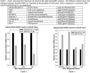
Table 1 lists seven Er-b-regulated genes expressed in LNCaP cells. Figures 1 and 2 show the relative steady-state mRNA levels of all seven genes in cells treated with different levels of Se. The expression of three genes: cdk2, pdl1, and gal1, was downregulated at high Se treatment, as shown in Figure 1. The expression of two genes, her-2 and ctsD, was unaffected by high Se treatment, as shown in Figure 2. Surprisingly, the expression of two genes, IGFBP10 and ha-ras was upregulated by high Se treatment.
Conclusions
Decreased steady-state mRNA levels for cdk2, pdl1, and gal1 are consistent with the hypothesis that Se acts by altering the conformation and activation of ER-b. The apparent upregulation of IGFBP10 and ha-ras expression by Se treatment, and absence of a significant effect of Se treatment on her-2 and ctsD, do not necessarily contradict this hypothesis. These results may be due to preferential regulation of these genes by transcription factors other than ER-b. Se-ER-b interaction is one possible mechanism for Se’s chemoprotective effect in prostate cancer.

