Daniel Tandberg and Dr. Merrill J. Christensen, NDFS Department
Introduction
Selenium has been established as a promising chemopreventive element for prostate cancer. Several different mechanisms have been studied to delineate selenium’s anti-cancer effect, but the exact target and mechanism has yet to be clearly defined. Previous studies have established an inhibitory relationship between the nuclear factor kappa B (NF-κB) and selenium, as well as the Androgen Receptor (AR) and selenium in human LNCap cells. Both NF-κB and AR are constituently active during prostate cancer development, and their deactivation by selenium might be one of the mechanisms by which selenium acts as a chemopreventative agent. The purpose of our research is to study the interaction between AR and NF-κB in the selenium pathway. More specifically, in this study we evaluated the effect of selenium in the form of methyselenilic acid (MSA) on the expression of AR and the subunits of NF-κB, p50 and p65.
Methods
Cell Culture: LNCaP human prostate cancer cells were derived from human lymph and present a good model of early androgen dependent prostate cancer. They have a mutant but functional androgen receptor. 22Rv1 cells have a functional androgen receptor but have been shown to grown independently to the presences of androgens. Thus, 22Rv1 cells can serve as a model of androgen independent prostate cancer. The LNCaP and 22Rv1 cells were obtained from American Type Culture Collection and maintained in RPMI 1640 supplemented with 10% heat-inactivated fetal bovine serum. The cells were supplemented with selenium in the form of methanoselenic acid (Sigma, St. Louis, MO). MSA is a biologically relevant form of selenium commonly used in cell culture. Confluent flasks were supplemented with 5 µM MSA 72 hours after being split. The incubation time was 72 hours, after which the cells were harvested.
RNA Isolation: Total RNA was isolated from cells using the Qiagen RNeasy kit (Qiagen) according to the manufactures protocol.
Real-Time PCR: After isolating the RNA, cDNA was synthesized using single base-anchored oligo dTs as previously described and as currently performed in the laboratory. AR, PSA, RelA, and p50 primers were designed using dsGene according to established protocols. Optimal annealing temperatures were determined by running a PCR temperature gradient on the RoboCycler. Real-Time PCR was performed using the Roche LightCycler with primer specific annealing temperatures. 18s rRNA was used to normalize final concentrations. Four runs of three replicates each was run with the LNCap cells. Two runs with three replicates each were run with the 22Rv1 cells.
Results
PCR Gradient: Optimal primer annealing temperatures were determined using a temperature gradient on the RoboCycler. The electrophoresis gel from RelA (p65) is pictured to the right as an example. 64 degrees was determined as the optimal annealing temperature for p65 based on the decreasing band strength at 66 degrees. The annealing temperature for the AR and p50 genes was 66 and 65 degrees, respectively.

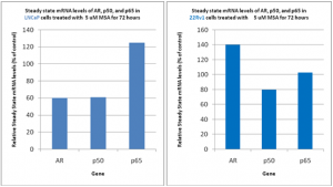
Real-Time PCR: 5 µM MSA reduced expression of AR and p50 in LNCaP cells after 72 hours of treatment, 60% and 62% respectively. Expression of p65 was enhanced. In 22Rv1 cells AR expression was enhanced, p50 inhibited, and p65 expression was not changed by treatment with selenium.

Discussion
Selenium disrupted gene expression for the androgen receptor and nuclear factor kappa-B in the LNCaP and 22Rv1 human prostate cancer cell lines. A subunit of NF-kB, p50, was inhibited in both cell lines which c9ould account for some of selenium’s anticancer effect. Further research is warranted to establish why selenium enhanced expression of AR in the 22Rv1 cells while inhibiting AR expression in the LNCaP cells.
