Jason Morris and Dr. Merrill J. Christensen – Nutrition, Dietetics, and Food Sciences
The goal of this project was to determine whether there was a synergistic anticarcinogenic effect between selenium and resveratrol, both of which had independently demonstrated the ability to inhibit prostate cancer cell proliferation. Additionally, the project also sought to determine whether a less soluble derivative of resveratrol, synthesized at BYU by Dr. Merrit B. Andrus of the Department of Chemistry and Biochemistry, maintained the anticarcinogenic potency of resveratrol, and what were the effects of this derivative in conjunction with selenium.
During this project two human prostate cancer cell lines were grown by standard cell culture methods in 25 cm2 flask. These two cell lines, LNCaP and 22Rv1, represented early and late stage prostate cancer respectively. These two cell lines were obtained from a reliable source, American Type Culture Collection. Cells were allowed to grow in the 25 cm2 flasks to approximately 80% confluency, at which time they were seeded onto a 96 well plate at a density of 5,000 cells per well for the LNCaP cell line and 10,000 cells per well for the 22Rv1 cell line. The cells were allowed to attach for 48 hrs before being treated with resveratrol, the resveratrol derivative, selenium, or a combination of these treatments for 72 hours. These protocols were determined by performing a cell titration assay which compared the absorbance of various cell seeding densities (1k-25k) and attachment times (24-48 hours) at a specified light wavelength that is indicative of cell proliferation. Once the optimal seeding density and attachment period were known we compared various treatment times (24-72 hours) to determine what length of time demonstrated cell proliferation inhibition that would not be attributed to lack of nutrient resources.
Having decided on optimal testing protocols we began testing different reagent concentrations to determine the effect on the proliferation of the cancer cells. We tested resveratrol and the resveratrol derivative at concentrations from 0-100 uM and determined that concentrations above 50 uM did not provide significant additional benefit. We had originally planned to test selenium concentrations from 0-20 uM, but after reviewing other studies and trials that were underway we narrowed our target concentrations to just 0-3 uM because levels greater than this were physiologically unattainable. In our original proposal we planned to measure cell viability in both cell lines using the MTS assay which measures mitochondrial activity. We also optimized this protocol by running tests on our plate reader to determine the optimal scanning parameters (7×7 matrix with 30 flashes). Because these optimized parameters resulted in significantly longer scanning times, we implemented a final step in the MTS assay that halted the reaction after one hour of incubation, providing us with a 24 hour window in which to scan the plate.
Originally we had planned to use the MTS assay to determine cell viability in both cells lines, but the high cost of the MTS assay led us to research other cell viability assays that we could use in our experiments. We determined that the crystal violet assay, a protein staining assay, could provided the same results at a fraction of the cost of the MTS assay. By replacing the MTS assay with the crystal violet assay we were able to reduce our assay costs per plate from approximately $40 to just a few cents. The crystal violet assay protocols proved to be a good fit for the heartier 22Rv1 cell line, but after a few failed attempts it was determined that the more susceptible LNCaP cells could not survive the rigors of the crystal violet assay intact.
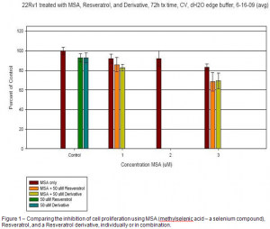
After refining and optimizing these protocols we were able to confirm that selenium and resveratrol inhibit carcinogenic cell proliferation, and that the novel resveratrol derivative also inhibited carcinogenic cell proliferation at levels comparable to resveratrol. Our initial data show that the effects of selenium and resveratrol may be additive, if not synergistic, but the data must be subjected to further statistical analysis before any such claims could be substantiated (Fig 1).
Once these findings are substantiated the next step will be to use cell biology techniques such as western blotting to identify the mechanisms whereby these agents exert their anticarcinogenic effect.

