Emily Brown and Dr. Dennis Shiozawa, Biology Department
Introduction
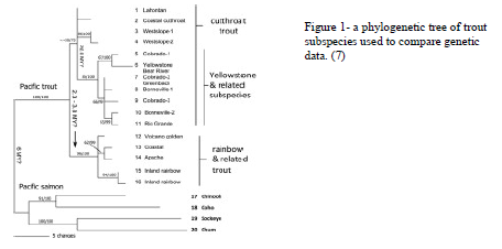
Since the 1800s, biologists have studied cutthroat trout native to Western North America. Their early work and classification were based on the standards of the day: meristics, the observation and counting of physical features, and morphology. Further improvements came through later studies that added geographic distribution to their phylogenetic classification. Without the foresight of DNA genotyping methods, “traditional taxonomic assessments often failed to accurately capture phylogenetic diversity.” Many discrepancies have since arisen as genetic methods shed new light on cutthroat trout subspecies phylogenies. Current mtDNA work has identified errors in historical classifications of several of these subspecies in the Colorado, Utah, Wyoming, and Idaho regions. Our new knowledge of misclassification has subsequently identified problems in protecting the right endangered subspecies. Genetic diversity progresses as trout populations change due to “habitat fragmentation, overharvest, introduction of nonnative species”, and other factors like sport stocking. DNA genotyping is crucial to protect these species from possible endangerment. Until recently genetic methods for classification have been limited to mtDNA, which utilizes a single locus, so developing a more comprehensive understanding of the nuclear genome is of great importance. The collaboration of the Kauwe lab and the Shiozawa lab at BYU, along with labs at the University of San Francisco, have almost completed sequencing the entire genome of one subspecies of cutthroat trout. Though an important accomplishment, more distantly related cutthroat trout are known to have a different number of chromosomes, so in order to maximize the usefulness of this newly defined genome, we need at least one additional completed genome from a related subspecies. Having completed genome scaffolds from two related subspecies will facilitate accelerated analysis and assessment of all the genetic relationships of cutthroat trout. We further believe that this approach can be adopted to the study of other species to more effectively protect them from endangerment and extinction. Our studies focused on how to develop the protocols and procedures needed to sequence the genome.
Methodology
Using the QIAGEN MagAttract HMW DNA kit for sample and assay techniques, we isolated our high-molecular-weight DNA samples from whole blood. We initially began with trial runs of rainbow and brown trout (diploid organisms) before moving to the cutthroat trout (tetraploid) to verify that we were doing the procedure correctly. We began by adding the enzyme Proteinase K to our blood sample, together with RNase A (a nuclease catalyst important to degradation of RNA) and Buffer AL solution to begin the lysis of blood cells. The solution was incubated during the lysing process. Then magnetic suspension beads were added, which bound to the DNA and were separated with a magnet. The supernatant was removed, product rinsed several times, and the suspension beads were removed, leaving the DNA product. We then ran the product on an agarose gel through electrophoresis. Once we had the DNA isolated, we used IrysPrep to label restriction sites in the sequences for complete assembly of the genome.
Results
This technique is new to our lab and took several weeks to troubleshoot. We made our own adjustments to the protocol after encountering problems such as coagulation, inactivity of the enzyme and improper lysis of blood. In our first few trials, we experienced issues with some of the provided materials from the QIAGEN kit. To start, the enzyme provided (Proteinase K) seemed to be inactive as it only resulted in mixed blood and coagulation. We believed this was potentially due to the enzyme being kept at room temperature, where it is normally stored at 5ºC. We opted to use our own enzyme from the cold storage, which seemed to solve the problem. However once that was resolved, we noticed the blood again coagulating after we added RNase A and Buffer AL solution to the sample. Through trial and error, we discovered that the buffer needed to be added first, (even though it was written as second in the protocol), and that the solution needed to be vortexed immediately after adding the RNase A to ensure a homogeneous lysed solution. Once these steps were perfected, we changed the incubation time to 50ºC for 45 minutes. This change came after researching prime incubation temperatures for DNA lysis. When it came time to separate the DNA product from the solute, the magnetic rack provided did not thoroughly separate the magnetic beads as efficiently as the magnet we already had at the lab. However, we resolved these issues and successfully isolated DNA product that we read on agarose gels. Unfortunately, because we initially ran these trial runs with our diploid samples from the brown and rainbow trout, we did not take pictures of the successful gel runs. However, we feel confident that using the MagAttract protocol for DNA isolation proved successful for diploid organisms.
Discussion
The complications we encountered resulted in high expenses and time lost in our efforts to move forward with this project. We submitted two runs to the lab in San Francisco for them to assemble and compare with our data. They had to assemble those runs several times, and it took us multiple attempts to download their data files. Due to entire genomes having tens of thousands of base pairs, it included so much data that each download required about 3 weeks, all of which cost us 5 months. The San Francisco lab also had unsuccessful attempts with their own DNA sequences and data analysis. We experienced other problems in assembling the rainbow and brown trout from our initial trial runs. If we had decided to move forward with the cutthroat trout, the assembly would have been even more complex, as salmonids are tetraploid organisms, leading to potential duplicate regions on different chromosomes.
Conclusion
After exploring the MagAttract and IrysPrep assembly procedures, we faced a crossroad to either continue down that path, or instead shift to a more standard technique with the PacBio. Despite successful DNA isolations from the MagAttract protocol, we had too many problems with technology downstream. We decided that while the procedure could now be done in-house, the limitation of the Irys technology meant that we would need to move to a more expensive sequencing platform, PacBio, which yields similar results from frozen tissue samples. We will now move forward using the PacBio to assemble our data and further pursue the research associated with this venture.

