Robert Fender and Faculty Mentor: Merrill Christensen, Nutrition, Dietetics, Food Science
Introduction
It is estimated that over 6 million Americans suffer from the neurodegenerative diseases of Alzheimer’s and Parkinson’s combined, with 1 in 4 Americans suffering from some type of mental illness (1). Oxidative damage and inflammation, which is a recognized hallmark of neurodegenerative diseases, can be prevented by antioxidant enzymes such as glutathione peroxidase whose key component is selenium and which relies on a selenium delivering protein called selenoprotein P (2). Since neurodegeneration that affects so many people can potentially be avoided by understanding how selenium is accumulated in and delivered to body tissue, understanding how and where selenium is accumulated in body tissue is of great importance.
Methodology
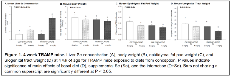
The purpose of this project was to gather further data on a study conducted previously by Dr. Christensen and his team regarding the relationship between dietary soy and selenium status in mice on one of two basal diets. One group was fed a diet of soy meal, the other was not. Within the two groups, half were supplemented with selenium while the other half was not. Therefore, there were a total of four groups. One which received soy and supplemental selenium, one which received soy but not selenium, one which received no soy but received selenium, and one which received no soy or selenium. Their question was whether or not soy affected the metabolism of selenium in a way that led to more selenium being retained in liver tissue. Data was collected which showed that mice fed a diet containing soy retained more selenium in their livers than the mice which were fed a diet with no soy (3) (Figure 1.4). The object of this study was to see if the same trend is true for their brain tissue.
This experiment employed the fluorometric assay developed by Koh and Benson for analysis of selenium. This method had been used in the previous experiment mentioned to determine selenium concentration in the mice livers (4).
Before the digestion and subsequent analysis of our sample tissues, we established a standard curve using NIST 1577c bovine liver standard reference material.
Results
As we began the tissue analysis with test samples, we realized that employing the flourometric method of analysis was not going to be practical due to the variability that could be introduced into the procedure. Upon a search into the current scholarly literature on trace element analysis, we decided to use microwave digestion together with inductively coupled plasma mass spectrometry to analyze our samples. We coordinated with Rachel Buck of the Environmental Analytical Laboratory at BYU together with Dr. Paul Farnsworth in the Chemistry department to develop a procedure whereby we could analyze a large quantity of samples free of the inconsistency found in the flourometric method.
Initial data supports the new method of analysis in that we can detect selenium levels within .5 part per billion increments and with a percent error of 1-2%. This is significantly more reliable data than anything we were able to achieve with the flourometric method. As a result of our developing this new analyzation method, we have been able to begin analysis of the brain samples and collect initial data.
Discussion
Although far from conclusive, our initial data from brain samples suggests that selenium is found at a much higher concentration in the brain tissue than in the liver. If this continues to be the trend, we may be able to make progress in our understanding the relationship between selenium status, diet, and neurodegenerative disease.
Conclusion
The major headway made in this study was that we were able to come up with a form of trace element analysis that is both efficient and extremely accurate. As a result, we have been able to begin analysis of the stored brain tissues with confidence that we are using the most current technology to get the most accurate and precise results possible. Recognition and thanks to the BYU ORCA program and it’s donors.
Sources
1. By the Numbers | McGovern Institute for Brain Research at MIT. (2014, January 16). McGovern Institute for Brain Research at MIT. Retrieved October 27, 2016.
2. Valentine, William M. et al. “Neurodegeneration In Mice Resulting From Loss Of Functional Selenoprotein P Or Its Receptor Apolipoprotein E Receptor 2”. J Neuropathol Exp Neurol 67.1 (2008): 68-77. Web.
3. Nakken, H. L., Lephart, E. D., Hopkins, T. J., Shaw, B., Urie, P. M. and Christensen, M. J. (2016), Prenatal exposure to soy and selenium reduces prostate cancer risk factors in TRAMP mice more than exposure beginning at six weeks. Prostate, 76: 588–596. doi:10.1002/pros.23150
4. Christensen MJ, Bown JW, Lei LI. The effect of income on selenium intake and status in Utah County, Utah. J Am Coll Nutr 1988;7:155–167.
