Jakob Gamboa and Dr. Jonathan Wisco, Physiology and Developmental Biology
In 1974, the first ulnar collateral ligament reconstructive surgery was performed on Los Angeles Dodgers’ pitcher, Tommy John. The damaged ligament of the medial elbow was repaired with tendons from the pitcher’s body. Since then, the surgery has been colloquially termed “Tommy John’s Surgery”, and the alarming increase of the rates of the procedure has now become a concern, being recently called an “epidemic” by the American Sports Medicine Institute1. The procedure possesses risk of complications, and can lead to shortened careers, decreased performance over time2, and can carry a heavy financial toll on individuals and athletic programs during the rehabilitation process.
Injury to the UCL can result from deterioration from the repetitive stress of overhand throwing motions, or can be caused by acute rupture from a single traumatic episode. The combination of force and velocity causes micro-tears and attenuation which eventually leads to failure of the fiber bundles. The UCL is composed of three ligaments, or bundles, with distinct fiber orientations: the anterior, posterior, and transverse bundles. The anterior bundle (AB) is the most commonly injured, and is the primary restraint to valgus forces, being subject to near-failure stresses during the extension of the elbow3.
Upon analyzing ten cadaveric ligaments to better characterize the mechanism of UCL injury, we discovered an anatomical variation with transverse bundle. The purpose of this study is to describe the previously undescribed variation and the clinical implications for greater stability with high performing athletes, and improved rehabilitation for Tommy John’s surgery (TJS).
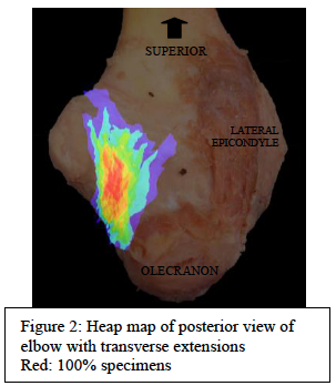
After collaborations with UCLA and West Virginia University, thirty three cadaveric elbows from 22 individuals were dissected and analyzed for insight regarding the prevalence and morphology of the anatomical variation. Included in the sample, were 20 right elbows and 13 elbows from the left limb. A deep dissection was performed to remove skin, fascia, and muscle tissue and tendons to reveal the ligaments of the medial elbow. The fibers were photographed in high resolution and compiled into a heat map in Adobe Photoshop to display variation.
Dissection of the cadaveric elbows revealed previously undescribed anatomical variations in 59% of the individuals. Twenty-one elbows from 13 individuals possessed ligamentous tissue, posterior to the ulnar collateral ligament. The fibers branch from the transverse ligament and attach posteriorly, beyond the typical attachment at the medial aspect of the olecranon, adjacent to the medial epicondyle. Due to this relation, we will refer to this posterior branch as the transverse extension (TE). The TE fibers extend an average of 30.5 mm ± 4.0 mm beyond the olecranon to the medial posterior elbow and/or medial supracondylar ridge, displaying deviating superior attachments. The supposed transverse extension (TE) variant exhibits individual fiber diameters of .2 ± .05 mm, analogous to the corresponding transverse ligament. The AB, PB, and TL are present in all specimens with typical fiber orientations, with the posterior bundle positioned deep to the transverse ligament and transverse ligament extension.
The presence of additional connective tissue on the elbow may reveal increased joint stability on the medial elbow. Continuous fibers from the medial transverse ligament to the posterior elbow offer additional attachment and expand a greater distance across the joint to improve function of this UCL bundle. The TE displays a similar vector to the AB across the elbow on the posterior region of the elbow, and based solely on the geometry of the fiber band, may function as an additional medial restraint to support the AB against valgus stress. The presence of such a variant would provide greater stability during overhand throwing motions to reduce overall risk of injury of the UCL. However, if the TE fibers are disadvantageous, then we assume that athletes would possess a greater risk of injury with its presence. If the greater resistance by the extended ligament tissue resulted in greater strain to the anterior bundle, we could predict that athletes who experience UCL tears and undergo TJS may likely possess the TE variation. In this case, the localized tissue could alter pre-surgical planning and outcome. During the reconstruction of the UCL, this additional fiber band could become a candidate for replacement tissue.
Analysis of the ulnar collateral ligament of 33 cadaveric elbows reveals anatomical variations of the transverse bundle in 21 of the specimens, representing 64% of sample specimens. The additional ligament fibers were discovered on the posterior elbow, continuous with the transverse bundle. The extension of the transverse ligament, with posterior attachments beyond the olecranon, has never before been described in literature. Clinical implications were proposed regarding the function and surgical uses of the ligament variant. Based on position, we discuss the possibility of providing restraint to valgus stress and additional medial stabilization to reduce injury of the anterior bundle and UCL complex, and surgical implications of such tissue for the prevalent Tommy John’s surgery.
- “Position Statement for Tommy John Injuries in Baseball Pitchers.” American Sports Medicine Institute. N.p., July 2014. Web. Nov. 2014.
- Makhni, Eric C., MD. “Performance, Return to Competition, and Reinjury After Tommy John Surgery in Major League Baseball Pitchers.” The American Journal of Sports Medicine, 4 Apr. 2014. Web. Feb. 2014.
- Cain, E. L., Jr. “Elbow Injuries in Throwing Athletes: A Current Concepts Review.” American Journal of Sports Medicine 31 (2003): 621-35. Print. Figure 1: Fiber bundles of ulnar collateral ligament http://jbjs.org/content/94/8/e49

