
Nathaniel Hainsworth and Jon Wisco, Physiology and Developmental Biology
Introduction
Not all tendons and ligaments are created equally. Many have different structural characteristics based on functionality or need for stability. This turns out to be true even between different portions of the same tendon. This project’s purpose was to analyze and compare structural differences between tendon fibers of the patellar portion and quadriceps portion of the quadriceps femoris tendon. We hypothesized that there are significant structural differences between these two portions because they act very different biomechanically, as well as different in the amount of tension/strain put on each portion. We mapped individual tendon fibers of both portions using MicroScribe technology, which gave us more detailed data about fiber patterns and structural differences including spacing, width, etc.
Methodology
Digital Imaging has been used in research with many tissues including nerve mapping, precise muscle attachment imaging and tendon differentiation. Most of these imaging techniques involve MRI technology. This project used a less common, and potentially more precise digital imaging technique (MicroScribe) that tracks and images individual fibers throughout the entire tendon. Using this method, we were able to map and calculate specific tendon differences, which will be described below. The actual MicroScribe technology is a tool that traces each fiber to produce a 3D rendering of the individual tendon fiber, that when done hundreds and hundreds of times, created a virtual tendon in an animation program. These digital recreations of tendon fibers are valuable because the fibers are easily viewed without connective tissue, fat, or vasculature between the fibers. We specifically dissected a sample from the quadriceps portion and patellar portion, which were then segmented coronally so that they were one fiber thick. These segments were then stained and MicroScribed into an animation program, Maya Autodesk. Once all layers were digitized, the layers were combined to create the digital tendon.
Results
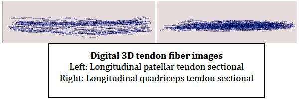
This study used these methods described above to get data from 5 cadaveric specimens, three embalmed, and two un-embalmed fresh specimens. Initial results demonstrated an increased number of tendon fibers accounted for than previously assumed, presumably due to the stain which accentuated the fibers and made them much more visible. Also neither tendon segment demonstrated any fiber crossing over, which is characteristic in many large tendons including the calcaneal tendon. In comparison between the two portions, the quadriceps portion was shown to run more obliquely in the sagittal plane, usually right as the quadriceps muscle body ends (proximal end). The patellar portion was shown to run almost completely vertical until the last few millimeters in which is splays out prior to connection with the bone (distal end). Although the patellar portion and quadriceps portion are technically part of the same tendon, The individual fibers in the patellar portion were shown to be thicker than the fibers of the quadriceps portion. Also the patellar fibers were more spaced out from each other than the quadriceps portion. Images of the MicroScribed tendon fibers are shown below.
Discussion
The major benefit of this research will be additional knowledge and information into the deeper makeup of these tendon fibers. Knowing spacing between fibers, width of fibers, and direction/crossing patterns of fibers will hopefully benefit in surgical and other medical procedures. The best example of this is ligament reconstruction surgery. Currently, many ACL repairs are reconstructed from borrowed calcaneal tendon from the ankle. This poses some risk because these tendon fibers differ in their crossing patterns and tendon width from the ACL. With this MicroScribe data, surgeons may be able to match tendons more precisely when deciding where to borrow tendon for reconstruction. Additional research is needed to branch into many more tendons/ligaments (ACL, PCL, Deltoid, biceps, triceps) to look for patterns or similarities between different major tendons. This bank of tendon information can be used to personalize reconstructive surgery choices with individual needs or injuries.
Conclusion
Most of us think of all muscles being the same and all tendons as being equal. Tendons are far from equal. They differ in size, shape, fiber width, space between fibers etc. This research dove into the differences between just two of the hundreds of tendons in the human body. This research is a culmination of my passion for the human body, knees in particular, which drove my investigation into the tendons of the knees and how they worked differed. In Dr Jon Wisco’s lab, we started MicroScribing bones and muscles, and we were able to expand that to tendons. Through MicroScribe technology we were able to find significant differences in tendon fibers, in addition to creating a new stain in which enhances the fibers to become more visible and easily distinguished. This ORCA grand provided funds to obtain cadaveric specimens as well as travel to the American Association of Clinical Anatomist’s annual conference in which this research was presented and published.