
Hannah Waddel and Dr. Arminda Suli, Physiology and Developmental Biology
A major task of the central nervous system is to make sense of the environment and mount an appropriate reaction. The ability to organize and integrate inputs from different sensory systems is critical for survival in animals. Sensory integration dysfunction is implicated in human disorders such as autism and dyslexia. Swimming behaviors such as rheotaxis, defined as swimming against a current, require the integration of different sensory systems such as mechanosensory hair cells, the visual system, and the vestibular system. In this project, we attempted to create an assay to measure the rheotaxis behavior of Danio rerio (zebrafish) larvae, in order to detect mutations in sensory integration systems.
We designed and constructed the assay, which contained ten separate lanes for larvae. Embryo medium was pumped through the apparatus with a peristaltic pump. We determined the flow rate using phenol red dye and adjusted it to be 0.2 cm/s, based on Dr. Suli’s previous research in rheotaxis (1). We placed one larva per lane and initiated a flow, and took one minute videos at 24 fps using a Point Grey Flea3 camera. The camera, apparatus, and pump were placed in a dark cabinet (Figure 1). We illuminated the apparatus from beneath using a light box.
When we had obtained the videos, we analyzed them using ImageJ video analysis software. Using a custom JavaScript macro, we collected each larvae’s X coordinate, Y coordinate, and head angle (Figure 2). We used the R programming language to clean the data and determine mean head angle for each fish during rheotaxis. The mean head angles for each group were compared using a one-sided t-test.
To determine the effect of the lateral line input on rheotaxis, I ablated the lateral line input by treating the larvae with 400uM neomycin. I completed five trials of neomycin-treated and wild-type fish. In the first trial, I obtained data from 20 wild type larvae and 13 neomycin-treated larvae. In a t-test of significance for the difference in mean head angles, the p-value was 0.155. In a second trial, with 16 wild-type fish and 18 neomycin-treated fish, we obtained a p-value of 0.078, which is not significant at the p=0.05 level. In the next trial, with data from 24 wild-type and 23 neomycin-treated larvae, the p-value for the difference in mean head angle was 0.007. In a final trial, with 7 wild-type fish and 7 neomycin fish, we obtained a p-value of 0.025.
To determine the effect of the visual input on rheotaxis, I tested the behavior in the dark, using an infrared array for illumination. With data obtained from 13 wild-type fish and 18 fish in darkness, we obtained a p-value of 0.021 for the difference in mean head angle. To ablate the vestibular input, I planned to treat transgenic embryos that express the nitroredactase gene in the eighth nerve, which is a gene that has a cytotoxic effect on the cell upon the addition of metronidazole (MTZ), and then test rheotaxis. To determine if MTZ alone affects the behavior of wild type larvae I treated groups of 30 fish with MTZ at a concentration of either 10 mM, 5 mM, or 1 mM. The fish that were treated with 10 mM MTZ all died. When I treated a second group with 10 mM MTZ, only 8 survived, and they did not swim at all in the assay instead laying on the bottom or near the edges. The group treated with 5 mM MTZ had a similar survival problem. Only 7 survived, and they did not swim in the assay. In the group treated with 1 mM MTZ, when we compared mean head angle between 9 treated fish and 17 wild type fish, we obtained a p-value of 0.248. While this high p-value indicates that MTZ at 1 mM has no significant effect on head angle, at that low concentration it did not properly destroy the cells in the eighth nerve. As a result, we could not test the effect of vestibular input on rheotaxis.
These results suggest that hair cells and visual input have an effect on a larva’s rheotaxis behavior. However, we faced difficulty in obtaining data from all of the fish that we tested with the assay. Due to friction at the sides of the lanes, the embryo medium flows at a slower rate, creating a place where larvae can avoid most of the force of the current. Of the wild-type fish, 52.9% of them were unable to be analyzed for swimming behavior because they used this boundary layer effect. 58.9% of the neomycin-treated fish and 40% of the fish in infrared light behaved the same way. Assuming that we were able to detect a mutation with at least 10 mutants, in order to detect a mutation we would need to run at least 98 larvae to find the expected 25% recessive mutants among the expected 41.1% of mutants we are able to film.
In conclusion, our assay is not sensitive enough to determine mutants. Tests of significance produced varying results, though the majority of the tests we conducted suggested that hair cells and visual input help determine rheotaxis behavior. However, due to the boundary layer effect at the sides of the lanes, we need an unreasonable amount of larvae to detect mutants. Though our assay could not determine mutants using rheotaxis behavior in a flow, our camera configuration and the video analysis macro we made will be useful for other behavioral assays.
References:
1. Suli A, Watson GM, Rubel EW, Raible DW. Rheotaxis in Larval Zebrafish Is Mediated by Lateral Line Mechanosensory Hair Cells. Riley B, ed. PLoS ONE. 2012;7(2):e29727. doi:10.1371/journal.pone.0029727.
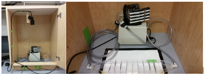
Figure 1. Configuration of our assay. The peristaltic pump moves embryo medium through the assay while the camera films.
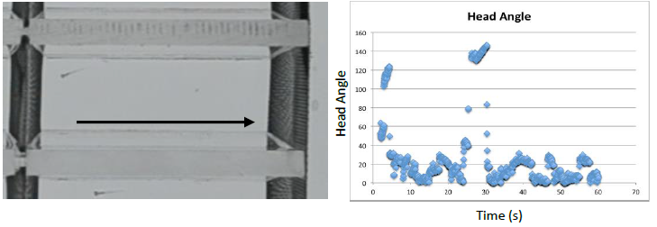
Figure 2. A fish engaging in rheotaxis behavior. The current flows in the direction of the arrow. Head angle of zero indicates rheotaxis (alignment with the current)