Dani Peterson and Jonathan Wisco, PDBio
Introduction
Due to its long and complicated trajectory through the cranium, facial nerve VII (CN VII) (see figure 1) can be damaged in surgeries, sometimes resulting in facial muscle paralysis. Surgical removal of acoustic neuromas and parotid tumors, in addition to surgical repair of the temporomandibular joint disorder are associated with a risk of damage to CN VII. In addition, insertion of auditory implants can damage the nerve, as can improper stimulation to the nerve after the implantation has occurred. This study’s focus was the creation of a three-dimensional (3D) model based off of data from dissection of the nerve in a human cadaver in order to give physicians a greater in vivo knowledge of the pathway of CN VII thereby decreasing the risk of damaging the nerve. Hypothesis: We hypothesize that with our approach and MicroScribe technique, we will be successful in creating an accurate model of CN VII.
Methodology
Dissection of Facial Nerve VII:
Two cadaveric heads embalmed in paraformaldehyde were acquired from the University of Utah’s Body Donor Program. Following bisection of one head, we dissected the lateral side of the right half of the head to the level of the parotid gland and identified the parotid plexus of CN VII. We identified and followed each of CN VII’s five branches. In addition, we followed the nerve through the internal auditory meatus on its pathway through the temporal bone. After bisection of the second head, we followed the nerve as it exits the brainstem and identified the greater petrosal nerve and chorda tympani.
Modeling Technique:
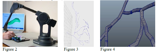
We tracked the nerve trajectory using a MicroScribe 3D Digitizer (see figure 2). The MicroScribe technique is used to create 3D computer models of any physical object. The user sets reference points and then uses the stylus to trace data points of the object’s contours at every 2-5 millimeters (see figure 3). This data was imported into the Autodesk Maya 3D digitization program, and the MicroScribe plotted points were translated into cylinder-shaped nerve representations (see figure 4).

Results
We demonstrated the ability to create a data-driven model of CN VII using the MicroScribe technique to plot the nerve’s trajectory and the Maya program to create its cylindrical representation. The resulting models detail the branching of CN VII into different planes of the face (figure 5) and the nerve as it exits the brainstem. In addition, we were successful in superimposing the nerve models onto representations of the cadaveric specimen constructed using MicroScribe (see figure 6) as well as the actual specimen (see figures 7, 8, and 9).

Discussion & Conclusion
This model provides a visualization of CN VII in space and demonstrates the presence of branches traveling in different facial planes. This finding leads us to suggest that facial muscles may be arranged in compartments. Thus, as the nerve branches exit the parotid gland, they may be destined for a specific compartment, explaining the planar variability. Following the reconstruction of the complete CN VII, this project will continue with comparisons of our in vivo nerve model with atlas recreations as seen in figure 1. Specific applications of this model to prevent potential injuries during removal of acoustic neuromas and parotid tumors, temporomandibular joint repairs, and auditory implant insertions exist as clinical goals.
