
Lauren Richey and Dr. John Gardner, College of Life Sciences
Main Text
A photonic crystal is a periodic structure that confines, manipulates, and guides photons. Such structures affect the propagation of electromagnetic waves by defining allowed and forbidden frequency bands known as photonic band gaps. Therefore, a photonic crystal can be thought of as the optical counterpart of a semiconductor, allowing only certain frequencies, or wavelengths, to propagate through the crystal. It is difficult to create photonic crystals operating in the visible frequencies due to the requirement of small periodicities to interact with visible light frequencies.
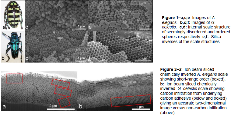
The structures that cause insect iridescence have periodicities with the correct dimensions to be photonic crystals. The structures of iridescent beetle scales of Glenea celestis and Anoplophora elegans (Figure 1a,b) in the Cerambycidae family were analyzed to probe their photonic crystal properties. Furthermore, inverses were created to test their possible photonic band gap. This beetle family, in contrast to the diamond structure of the Curculionidae family, has an array of tiny spheres inside their iridescent scales which resemble mineral opals and colloidal sphere arrangements which are commonly used by researchers to create “inverse opal” photonic crystals.
The internal scale structures of G. celestis and A. elegans were viewed with a scanning electron microscope (Figure 1c,d), but these images were inadequate to fully understand the structure. Using a combination of focused ion-beam (FIB) milling and electron microscopy, two dimensional images of the beetle scales were obtained from the microscope to create a highresolution three-dimensional scale reconstruction through a “slice and view” technique. This method of slicing the biological beetle did not work as the spheres melted together creating messy two-dimensional images. Inorganic replicas of the scales were created through a chemical inversing technique. By infiltrating a mesoporous silica sol-gel between the spheres and then dissolving out the original spheres, an inverted silica lattice was created which was similar to the inverse opals researchers create from colloidal sphere arrangements (Figure 1e,f). Not only was an inverted structure of the Cerambycidae scales the correct type of structure for a photonic crystal, an inverted scale was also the easiest to work with using the FIB microscope.
By analyzing a combination of the three-dimensional reconstructions, the image stacks from the inverted scales, and the single images from the original biological scales, the arrangement of the spheres in G. celestis was found to be face-centered cubic. The structure of A. elegans is still the subject of investigation and may contain short-range hexagonal order or exhibit a quasi-periodic sphere arrangement (Figure 2a).
These results were presented at two different conferences. A poster was presented at the 2010 Microscopy and Microanalysis Conference in Portland, Oregon from August 1-5, receiving the second place student biology poster award and an invitation to write an article for Microscopy Today magazine (a work in progress). Another poster was presented at the APS Four Corners meeting from October 15-16, winning the “best paper” award.
Over the course of this research, there were many complications that revolved around the operating ability of the FIB microscope. Besides regular instrumentation problems (e.g. stage alignments and gallium ion beam source replacement), most of the summer was spent dealing with vibrations from construction on upper campus. These vibrations shook the microscope while delicate slice and view runs were in progress thus ruining many samples and forcing slice and view runs to be completed at night.
Furthermore, the creation of better three-dimensional reconstructions is a work in progress. As seen in Figure 2b, the image appears slightly blurred because rather than obtaining a true twodimensional view, the image shows other parts of the lattice that are behind the milled face thus allowing the repetition of information from slice to slice. However, an accurate two-dimensional slice can be seen at the bottom of Figure 2b with no additional three-dimensional information. This was determined to be a result of the underlying carbon adhesive (that holds the sample) being forced up into the inverted scale. This realization has led to the idea of purposefully infiltrating carbon into the silica lattice to obtain better two-dimensional images from the FIB microscope. This is a work in progress and will hopefully assist in the analysis of A. elegans and the improvement of the three-dimensional reconstructions of G. celestis.
The analysis of the possible band gaps of these structures is also underway and will be completed by January. By placing quantum dots in the crystal that radiate in the frequencies of the suspected band gap, one can “test” the band gap. In photonic crystals, the decay of an emitter, such as a quantum dot, is inhibited quite measurably within the band gap, whereas when the frequency is outside the band gap, its emission will be enhanced. This will help in the characterization of the band gaps in fabricated crystals.
Special Thanks
Special thanks to Michael Bartl and Mathew Jorgensen at the University of Utah and Michael Standing at Brigham Young University for helpful insights as well as David Belnap at Brigham Young University for help in the three-dimensional reconstruction.