Britlyn Orgill and Dr. Merrill Christensen, Department of Nutrition, Dietetics, & Food Science
Main Text
Prostate cancer is the second most common cancer in American men. The American Cancer Society predicts that 217,730 cases of prostate cancer will be diagnosed and that 32,050 men will die from the disease in the year 2010 (1). Since prostate cancer has a long latency period, it is a prime candidate for dietary prevention. Isoflavones, natural compounds found in soy, and selenium, a trace element, have both been previously shown to have protective effects against prostate cancer. The lab of Dr. Merrill Christensen has been studying the effects of these compounds using the Transgenic Adenocarcinoma of the Mouse Prostate (TRAMP) model.
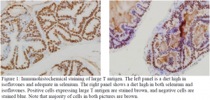
The TRAMP model uses the large and small T antigens of the Simian vacuolating virus 40 (SV40). The large T antigen causes tumorigenesis through inhibiting the p53 (tumor protein 53) and Rb (retinoblastoma protein) tumor suppressor genes. In the TRAMP mouse, expression of the large and small T antigens is directed to the prostate via the rat probasin promoter. The probasin promoter has two androgen response elements where androgens bind. This binding of androgens is necessary for the expression of the T antigens, and therefore tumorigenesis. The TRAMP model demonstrates aggressive prostate cancer as mice can present neoplasia as early as 10 weeks and well differentiated adenocarcinomas at 24 weeks (2). Our laboratory studies the effects of selenium and isoflavones on prostate cancer through four diets: adequate selenium and adequate isoflavones, high selenium and adequate isoflavones, adequate selenium and high isoflavones, high selenium and high isoflavones. However, since selenium and isoflavones have been shown to knock down androgen expression, it is possible that the diets are affecting the TRAMP model instead of the cancer. Since many proteins we investigate using TRAMP mice are androgen-dependent, we needed to test the TRAMP model. The purpose of this project was to determine whether there was an appreciable difference in large-T antigen expression among the four diets.
To look at expression of large T antigen, we used immunohistochemistry. This project introduced of immunohistochemical techniques to the Christensen lab. We used dorsal-lateral prostate tissue from mice sacrificed at 18 weeks as we have found this time point to show significant differences in expression of other proteins between the four diets. We looked at specimens from 5-6 animals per diet. After harvest, the tissues were formalin-fixed for 24 hours. The tissues were transferred to 70% ethanol and then processed and paraffin-embedded. Using a microtome, we cut 6 μm sections, which we then transferred to Superfrost Plus slides.
We developed an immunohistochemical protocol specific to our antibody and tissue that used a Horse Radish Peroxidase system and kit reagents (Dako North America, Inc., Carpinteria, CA). Endogenous peroxidases were quenched and large T primary antibody was applied at a 1:400 dilution in .2% Bovine Serum Albumin and incubated overnight at 4° C. Antigen retrieval was performed using 6 mM citrate in a water bath at a temperature 95-100 ° C. Biotinylated secondary antibody was applied followed by incubation with streptavidin. Positive cells were stained using 3,3′-Diaminobenzidine chromogen and a counterstain was applied using methyl green. Slides were coverslipped using Permount. Four to six pictures were taken per slide at 400x magnification using an Olympus microscope. Positive and negative cells were quantified using Image J software (NIH, Bethesda, MD) by two independent, blinded observers (Figure 1).
After several refinements of our protocol we were able to successfully quantify large T antigen expression across the four diets. We hope to publish these results in a peer-reviewed, scientific journal in the near future. In addition to learning more about the TRAMP model, this project also led to the introduction of immunohistochemistry to the Christensen lab. This procedure is now an asset that we can use to quantify proteins in addition to western blots. We have plans for this coming semester to use our immunohistochemical techniques to look at CD31 and Ki67 expression. These antibodies are markers for angiogenesis and cell proliferation respectively.

Acknowledgements
I would like to thank TJ Randall and Trevor Quiner for their contributions to this project. I would also like to thank Dr. Paul Reynolds for the use of his tissue processor and Dr. Robert Seegmiller for the use of his microscope. I also thank the Wound Healing and Tissue Engineering Laboratory at Brigham and Women’s Hospital, Harvard Medical School for teaching me basic immunohistochemical techniques.
References
- American Cancer Society. Prostate Cancer. www.cancer.org/Cancer/ProstateCancer/DetailedGuide/prostate-cancer-key-statistics. Nov 2010.
- Gingrich JR, Barrios RJ, Morton RA, Boyce BF, DeMayo FJ, Finegold MF, Angelopoulou R, Rosen JM, and Greenberg NM (1996). Metastatic Prostate Cancer in a Transgenic Mouse. Cancer Research, 56, 4096-4102.
