Ryan Martin and Professor David Busath, Physiology and Developmental Biology
Main Text
General anesthetics are widely used today in medicine. Their molecular mechanisms however, still remain a mystery. For centuries, general anesthetics were thought to be “drugs without receptors” because of the lack of knowledge about their mechanism of action (Hemmings, et al., 2005). While some advances have been made by Neuroscientists and Biologists there is still a lot that is not known about general anesthetics. The exact location of general anesthetic action is thought to be complex, but two particular locations have been suggested—the cerebral cortex and the midbrain. Detailed understanding of where general anesthetics act is important because it would allow doctors to better control anesthetic side effects, patient awareness during surgery, and fatal allergic reactions to anesthesia. Furthermore, enhanced understanding of general anesthetics could play an important role in better understanding the human consciousness. The better we understand general anesthetics, the better we will understand consciousness and vice versa.
The two hypothesized areas involved in consciousness are the cerebral cortex and the midbrain. Published research appears to support both hypotheses. The cerebral cortex is thought to be involved with consciousness because of its functions in executive skills and cognition. The midbrain is thought to potentially be involved because of its control of arousal states. Our goal with our project was to test one of these two hypotheses using slices of mouse brains to do tests in vitro. In order for our project to be a success my mentor and I collaborated with various professors. One principle collaborator was Dr. Scott Steffensen of the Psychology Department. Over the course of our project Dr. Steffensen provided expert knowledge on the electrophysiology of the mouse brain and gave us the necessary training to learn important in vitro techniques for our project.
Originally our hypothesis was to measure the amount of brain activity in the cerebral cortex of mice in vitro when stimulated with and without anesthetics. Before we got too far on the project Dr. Steffensen suggested that we switch our hypothesis from “the cerebral cortex as the effect site for general anesthetics,” to “the midbrain as the effect site for general anesthetics.” Because of Dr. Steffensen’s expertise in the midbrain, we trusted his advice and made the change.
As part of my project I learned many new laboratory skills such as how to prepare and dye brain tissue, how to accurately use a light microscope, and how to use imaging software. I also learned how to harvest mouse brains for in vitro research. We obtained mouse brains from female rats that were deeply anesthetized with isoflurane, and then decapitated; their brains were then removed, chilled and thinly sliced using a vibratome stage. These slices were then stained with the appropriate concentrations of dye and prepared for imaging.
Our research design involved using Voltage Sensitive Dye (VSD) and a fast speed imagining camera to measure brain activity in the prepared brain slices. In theory we would stimulate the tissue with an electrode and cause millions of neurons to fire. The dye in the tissue allowed us to be able to visualize the wave of action potentials as it moved along the cerebral cortex. The fast speed camera would catch the light and create an image. Our hypothesis was that by administering general anesthetics to the tissue in vitro we should see some sort of change in the light emitted by the tissue when compared to non-anesthetized tissue.
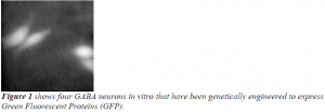
Imaging action potentials was a new task for all involved in the project, including the professors. We spent many months troubleshooting problems with camera lenses, filters, dyes, noise filtration, software programs, and more. The mice we used for imaging had been genetically engineered to express Green Fluorescent Proteins (GFP) in GABA neurons throughout the brain (see Figure 1 below). Glowing GABA neurons were used to help us troubleshoot problems with imaging software and hardware. In addition GABA neurons were used as landmarks when taking multiple images. Over the course of many months we learned a lot about imaging and in vitro studies, but ultimately we were not able to image propagating action potentials as we had hypothesized.

Our project was particularly difficult because of the skills we were required to master in a short period of time and the knowledge we were required to obtain from reliable internet databases. We did not save the world or publish a miraculous paper, but I gained experience that was invaluable. I will forever be grateful for my ORCA-sponsored research experience. I learned research can be very tedious and time-consuming, but it also can lead to great achievement and satisfaction. A special thanks is owed to my research mentor, Dr. David Busath, our collaborator Dr. Scott Steffensen, and all of the students that aided with their time and effort on the project. Although I am no longer involved, research continues in Dr. Scott Steffensen’s lab on a related project to the one I worked on. I wish them all the best and success in the future!
Sources
- Hemmings HC, Akabas MH, Goldstein PA, Trudell JR, Orser BA, Harrison NL. Emerging molecular mechanisms of general anesthetic action. TRENDS in Pharmacological Sciences 2005;26:503-10.
