Michael Henderson and Dr. Robert Seegmiller, Department of Physiology and Developmental Biology
The Need for a Mouse with Osteoarthritis
Osteoarthritis (OA) causes discomfort and immobility to millions of Americans every year. Due to the high demand for patient pain relief and improved mobility, scientists are seeking to understand the pathway of OA and develop drugs which would illuminate the onset of, and/or the progression of, OA.
Mouse models have previously provided an effective route for targeting the pathways of many diseases and discovering treatments. For OA, many mouse models have become obsolete or limited in their use, due to their dwarfed characteristics. For this reason we have put forth great effort in obtaining irrefutable evidence that establishes the Spondyloepiphyseal Congenita Heterozygous mouse (sedc/+) as the model scientists should use to study OA.
Two defining characteristics of the sedc/+ mouse validate its use in OA research. First, the arthritis that the sedc/+ mouse develops is due to a mutation in the C-Propeptide region of the col2a1 gene. This same mutation has been found to cause OA and other significant abnormalities in some humans. Because the sedc/+ mouse shares the same mutation as humans, its applicability to this human population will be readily accepted. Secondly, the sedc/+ mouse is non-dwarfed. Non-dwarfism validates the sedc/+ mouse to be used for studies applicable to the broader, non-dwarfed population of OA human patients. These two defining characteristics of the sedc/+ mouse establish a great need for a scientific study and establishment of the truth of these claims. The validation of the sedc/+ as a model for human OA should be published in a nationally recognized journal.
To establish the sedc/+ mouse as a valid model for OA research we set forth the following alternative hypothesis: sedc/+ mouse knee cartilage is significantly different (displays characteristics of early Arthritis) as compared to wild-type knee cartilage.
Establishing OA in the sedc/+ mouse
When I submitted my proposal for additional funding through ORCA, my team and I had been working on the sedc/+ project for about a year and had run into some difficulties. After receiving the ORCA grant we began to set goals once again and became determined to see the project through to completion. Our goals were to prepare a poster and present it at the Utah Conference of Undergraduate Research in February, conclude euthanizing and harvesting tissues (February), complete the cutting, or sectioning, of all the mouse knee joints (March), and write and submit a publication to Arthritis and Rheumatism Journal (May).
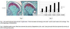
Three key steps were required to provide scientific evidence that the sedc/+ mouse develops OA faster than its wild-type (normal) counterpart: harvest, section, and analyze. First, harvest knee tissues of 4 mice at each age (2, 6, 9, and 12 months) for both sedc/+ and wild type. Second, embed knees in paraffin wax, section into slices 6 micrometers thick, place on small glass slides, and stain. Third, analyze observations of stained slides using the modified mankin score. Quantify results from analyzing and run statistical analyses with P <.05. The outlined steps were followed precisely despite difficulties encountered in determining the mouse genotype (sedc/+ or wild-type) and staining procedures. Staining created a great stumbling block to the progress of our project. To this day we are determining the exact problem. While we still have difficulties in this area, we were able to obtain enough coloring for our project to provide adequate results and proceed to analyze the slides. Sections of sedc/+ knee joints show an increase in red staining. This red staining provided evidence that the cartilages is weak and trying to rebuild. Therefore, sedc/+ knees show OA early in their life and this is shown by histological pictures. The mankin scores from our analyses provide substantial evidence of an increase in articular cartilage breakdown of the sedc/+ mice as compared to the wild-type. After our careful attention to detail and many revisions, the sedc/+ team consolidated the findings of our years of research and organized them into a publishable manuscript. On the 15th of October we received verification from the Arthritis and Rheumatism journal that they had received our publication and it was under review! Great excitement and feelings of accomplishment prevailed in the lab immediately following that notification.
Acknowledgements
- Office of Undergraduate Research grant and NIH grants
- Dr. Robert E. Seegmiller, Dave Holt, Davey McDonald, Jason Farrell, Steve Cowles, Josh Lloyd, Trenton Johnson, and Melissa Ricks

