Jillian Markham and Dr. Brian Poole, Microbiology and Molecular Biology
Background
Epstein-‐Barr Virus (EBV) causes infectious mononucleosis upon primary infection, commonly known as “mono.” Less commonly known is that EBV doesn’t get eradicated from your body after you recover from mono. EBV quietly occupies a small portion of B cells in 90% of human adults [1]. Besides mononucleosis, EBV infection is linked to cancers, lymphomas, and several autoimmune diseases [2.3].
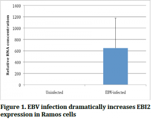
It was initially discovered that EBI2 (a host protein) expression was 200 fold higher in Ramos cells infected with EBV than Ramos cells with out EBV [4]. However, not much has been done since that study to figure out how and why EBI2 is so highly expressed in EBV infected cells. The goal of this project is to determine if there is a difference in EBI2 expression across different EBV latency stages in cell lines and in primary B cells in order to come closer to understanding the function of EBI2 expression in EBV infection.
Materials and Methods
The cell lines that were used were Ramos, Raji, and LCL. Ramos cells are naturally EBV-‐free, so they were used as the control. Raji cells are in latency stage 1 and LCL cells are in latency stage 3. Cell lines were grown for two weeks and then lysed and the RNA was extracted from them. The RNA was reverse transcribed into cDNA to be used in real time PCR to compare levels of expression of EBI2. Blood samples were also taken, and the B cells separated using a magnetic separation kit. The B cells from each blood sample were divided in half, with half of the B cells remaining as they were and the other half being infected with EBV. These cells were grown for one day and a sample was taken and at one week another sample was taken. All these samples also had RNA extraction, reverse transcription, and real time PCR performed along with the cell lines.
Results and Discussion
Our first experiment was to verify the previously mentioned finding that Ramos cells expressed EBI2 more when infected with EBI2. As is shown in Figure 1, we found this to be true, allowing us to use Ramos cells infected and uninfected as our controls.

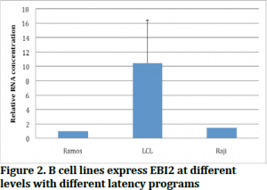
Figure 2 shows the results of our experiment with the different cell lines. We found EBI2 expression is indeed different between latency stages, specifically EBI2 is expressed higher in latency stage 1 than latency stage 3. This suggests that EBI2 might be important in the early stages of EBV infection, and that perhaps during infection EBI2 expression might become lower with time.

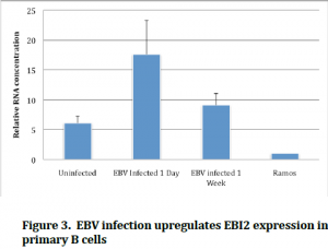
Figure 3 seems to confirm this, as we see that primary cells infected after one day have much more EBI2 then cells infected after one week. These are interesting findings because there has been no work done in primary cells with EBI2 before, and this gives researchers an idea of possible future directions to go with studying this gene. Now that we know what stages and times EBI2 is most highly expressed in, studies can be done with the genes that are known to be expressed in those stages. Understanding the infection patterns and genetics of EBV can help us to deal with the problems that are associated with EBV infection.

References:
- H.B. Warner, R.I. Carp, Multiple Sclerosis etiology — an Epstein-Barr virus hypothesis, Medical Hypotheses, 1988. 25(2): p. 93-‐97.
- Klein, G., et. al., An EBV-Genome-Negative Cell Line Established from an American Burkitt Lymphoma; Receptor Characteristics. EBV Infectibility and Permanent Conversion into EBV-Positive Sublines by in vitro Infection. Intervirology,1975. 5: p.319-‐334
- G. Pallesen, et. al., Expression of Epstein-Barr virus latent gene products in tumour cells of Hodgkin’s disease. The Lancet, 1991. 337(8737): p. 320-‐322. (http://www.sciencedirect.com/science/article/pii/014067369190943J)
- Rosenkilde, Mette M., et al., Molecular Pharmacological Phentotyping of EBI2: an orphan seven-transmembrance receptor with constitutive activity. Journal of Biological Chemistry, 2008. 281: p. 13199-‐13208.
