Derrick Crawford and Dr. Jeffery Barrow, Developmental Biology
Introduction:
Congenital limb malformations occur in approximately 1 in 500 live births. These defects are due largely because of genetic and developmental causes. In my experiments, I have researched the source of these defects to understand how cell to cell signaling affects digit and limb formation. I was able to manipulate signaling between limb tissues during development to understand better how proteins interact with each other to stimulate growth in chick embryos. These same proteins have been observed in mammals such as mice and hopefully in the future may be applied in human development as well. There are many key factors which play roles in limb development. One of these key signaling centers in the developing limb is the apical ectodermal ridge (AER). The AER is a ridge of tissue that is found at the distal margin of the embryonic limb bud. Past studies have revealed that when the AER is removed at early stages of development, the limb is severely truncated to the humerus. When removed at progressively later stages, increasing extents of distal development occurs (i.e., radius, ulna, and digits). It was proven later that members of the Fibroblast Growth Factor (FGF) family can replace the AER in signaling growth and patterning of the limb. My experiment was to study the effect of the FGF protein and it’s interaction with Sonic Hedge Hog, another protein involved in limb development.
Methodology:
In our experiment, we studied the effects of the protein FGF on developing limb buds. Using microsurgery, we opened up the embryo membranes and removed the AER from the distal tip of a chicken embryo incubated for 4 days. We then soaked microscopic beads in FGF protein and, using a tungsten wire, implanted them in the posterior end of the developing limb bud. (Figure1) After four days of incubation, living chicks were harvested and prepped in solutions for four days in order to clear tissue and stain the skeletal structure. We then observed and measured skeletal growth according to the differing concentration of FGF protein and the location of implantation.
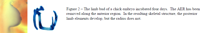
In the second half of our experiment, instead of completely removing the AER and replacing it with FGF soaked beads, we decided that we could obtain more accurate growth data if we staged experiments where varying amounts of AER were removed, leaving pieces in certain quadrants, and then observing the resulting growth. (Figure 2)
Embryos from each experiment were also stained for Wnt5A expression, a protein signal downstream of the FGF protein signal pathway. By measuring where Wnt5A was expressed, we could properly observe FGF penetration. (Figure 1)
Results/Discussion:
By surgically removing the AER during early stages of development and implanting the beads, we saw rescue of the truncated limbs. However, we found that the location of the implanted bead is just as important as the bead itself. We found that beads implanted posteriorly maintained much better rescue than bead implanted anteriorly. However in both cases, we observed very inconsistent results. In some cases we saw almost perfect rescue of the limb whereas in others, complete truncation still occurred because the bead was implanted improperly and fell out. After repeating the experiment multiple times with similar results, we looked to the Wnt5A expression stains and found that one of the reasons for the lack of resolution was that implanted beads caused expression of Wnt5A throughout the entire limb rather than in the area directly surrounding the bead. Therefore we could not make any accurate conclusions based on the location of the source of FGF.
We decided that instead of using beads to disperse FGF, we would use the AER itself. This time, rather than removing the entire AER, we would remove certain segments, and observe the resulting skeletal formation. Our results were many times more accurate and consistent. We found that when we removed anterior portions of the AER, the resulting anterior skeleton failed to form, but posterior segments, such as the ulna and digits grew to normal sizes. Interestingly, when the posterior region of AER was removed, complete truncation occurred. This is because of a small mass of cells located in the posterior position of the developing limb, called the Zone of Polarizing Activity (ZPA). These cells produce a signal called Sonic Hedge Hog (SHH) which we believe is another key factor in healthy limb development. Originally we planned on studying the effects of signaling between FGF and SHH, however because we were unable to obtain conclusive results from FGF soaked beads, we could not move on to SHH soaked beads. Instead we decided to gain a better understanding of the AER and its role in FGF secretion leading to limb development. These are key results for publication that we will submit in November.
Conclusion:
We have shown that each segment of the AER moving from anterior to posterior portions pertains to specific skeletal regions of development. We also concluded that though FGF soaked beads can cause partial limb rescue after AER removal, it is not able to replicate the physiological levels of secretion that partial AER removals reflect. Currently we are compiling our data in a paper that will be published in a reputable developmental biology paper. We have spent almost two years on our current project, and we plan on continuing our study of the AER and its crucial role in limb development. Because of the ORCA grant I was able to present our research at both the regional and international conference for the Society of Developmental Biology in Salt Lake City and in Cancun Mexico. Our poster received the finalist award among the 300 presenters that attended. As we continue researching in this field, we come closer to understanding limb development and the protein signals involved.
Sources:
- Niswander, L., C. Tickle, etal. (1993). “FGF-4 replaces the apical ectodermal ridge and directs outgrowth and patterning of the limb.” Cell 75(3): 579-587
- Wilkie, A. O. M. (2003), Why study human limb malformations?. Journal of Anatomy, 202: 27–35. doi: 10.1046/j.1469-7580.2003.00130.x
![]()