Scott Weber and Dr. Edwin Lephart, Zoology
Introduction:
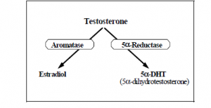
In certain brain regions, brain cell differentiation is influenced by hormones present at the time of development. Two hormones, estrogen (estradiol) and testosterone, play significant roles in male and female brain structure differentiation. The major androgen (testosterone) metabolizing enzymes, aromatase cytochrome P450 and 5a-reductase, play critical roles in the conversion of testosterone into estradiol and 5a-dihydrotestosterone, respectively.1
Phytoestrogens, naturally occurring compounds found in many foods, are defined as plant substances that are structurally or functionally similar to estradiol. Phytoestrogens bind the same sites as estradiol within cells and have similar effects as estradiol within the human body. Since phytoestrogens have similar effects as estradiol, phytoestrogens may affect brain development if an individual is exposed to high concentrations of phytoestrogens during central nervous system development. Phytoestrogens are found in large abundance in soy plants and soy products, including some human infant soy formulas. Various rat diets also contain phytoestrogens. In this study, brain aromatase and 5a-reductase, regulatory behaviors and testosterone levels were examined in adult rats on phytoestrogen diets.
Overview of the study
Adult (74 day-old) male Sprague-Dawley rats were randomly assigned to two different treatment groups (n=18). The phytoestrogens were administered in the following ways:
The male rats received phytoestrogens (via) solid and/or their liquid treatments (tap water or ProSobee human infant soy formula in their drinking bottles) for approximately three weeks. During the treatment period, daily food and water intake was measured. Also, open field behavior was measured at the beginning and end of the treatment interim to detect any potential differences in their ambulatory activity. At the end of the treatment interval, body weight and ventral prostate weight were measured. Trunk blood was collected for subsequent determination of testosterone steroid hormone levels via radioimmunoassay. Furthermore, the brain tissue sites [i.e., the medial basal hypothalamus and pre-optic area (MBH-POA) and amygdala area] were collected and assayed for aromatase and 5a-reductase enzyme activity levels by the titrated water release or thin layer chromatography methods.
Results:
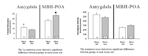
There were no significant differences between groups for food and water intake, body weight, open field activity, testosterone levels, and ventral prostate weights. Brain aromatase and 5a-reductase were measured in two separate tissue sites (MBH-POA and amygdala) as displayed below:
Significance
To date, alterations in 5a-reductase activity have not been observed even when rats were castrated.2 To our knowledge, this is the first time that brain 5a-reductase activity has been altered in a significant fashion based upon the presence or absence of phytoestrogens in the diets. The mechanism by which estrogen levels may affect 5a-reductase is not known. Further research is needed to understand the regulation of this pathway and the influence of phytoestrogen consumption. The data derived from this study is currently being prepared for submission to: The Proceedings of Experimental Biology and Medicine.
References
- Jacobson, N.A., Ladle, D.R. and Lephart, E.D. 1997. Aromatase cytochrome P450 and 5a-reductase in the amygdala and cortex of perinatal rats. Neuro Report 8: 2529-2533
- Lephart, E.D. 1993. A Review of Brain 5a-Reductase: Cellular, Enzymatic and Molecular Perspectives. Implications of Biological Function. Molecular and Cellular Neurosciences, 4: 473-4884


