Joseph Thurgood
SUMMARY
The palmar metacarpal aspect of 27 purebred (n=24) and appendix (n=3) American Quarter Horses were divided into sections and ultrasonically viewed to determine the circumference and cross-sectional area of the four principle tendons and ligaments in this region: superficial digital flexor tendon, deep digital flexor tendon, inferior check ligament, and suspensory ligament. This study was conducted using Brigham Young University Equitation schooling horses between May 1997 and February 1998. The structure measurements were analyzed for relationships to weight, age, sex and size of the horses (n=27). No correlation between weight, age, and sex of the horse and dimensions of any of the structures was found at any zone. No significant difference was found between measurements of the tendons and ligaments of left and right forelegs. These findings offer veterinarians a reference point to aid in diagnosis of forelimb injuries, such as tendon and ligament strains or tears. Hence, practitioners can more objectively evaluate the treatment response of an injured Quarter Horses by comparing the measurements of the injured leg to that of the normal leg.
INTRODUCTION
Ultrasound has become a useful diagnostic tool in many aspects of veterinary medicine. Recently ultrasonic techniques have been devised to more accurately diagnose equine lameness (1). Ultrasound allows one to quickly and non-invasively view soft tissue structures and facilitates differentiation and delineation of abnormal and normal size or echogenicity (2). The tendons and ligaments of the front metacarpal region of the horse are innately prone to injury and trauma during intense training and performance due to the horse’s anatomy. Through the use of ultrasound, tendon and ligament injuries can be more accurately diagnosed and the precise progress of an injury can be more readily assessed. Studies of this type, in general, have historically been performed to gain information on specific breeds (1). This study is of significant importance because it addresses the normal distal forelimb echogenicity of the breed that boasts the largest number of registered horses in the world, the American Quarter Horse. The objective of this study is to determine normal parameters for ligamentous and tendinous structures of the distal forelimb of the American Quarter Horse. This information should provide comparative values for practitioners evaluating forelimb lameness in this breed.
MATERIALS AND METHODS
The sampling group included Brigham Young University Equitation schooling horses of both sexes in the age range of 2-14 yrs. The horses were prepared for examination by walking them into restraint stocks, to assure safety to the horse and the examiner. Prior studies have shown that higher quality images can be achieved if the legs are shaved (3). Recently it has been shown that acceptable images can be achieved without removing all the hair (3). The legs were show clipped but not shaved to the skin. The caudal region of the metacarpal aspect was scrubbed with isopropyl alcohol starting at the distal point of the accessory carpal bone and extending distad to the fetlock joint. A 28 cm portion of medical tape was demarcated at four centimeter increments and placed on the lateral aspect of the metacarpal region, beginning at the distal point of the accessory carpal bone and extending distad to the fetlock joint (4).
An ultrasound machine (a) and a four centimeter ultrasonic standoff pad (b) were used for the examinations, both of which were provided by the Brigham Young University Animal Science Department. The machine was set to 7.5-MHz and a depth of six centimeters. The well lubricated standoff pad was placed over the ultrasonic transducer. Examination was performed on the caudal aspect of the metacarpal region of each foreleg. Depending upon the length of the cannon bone, there were a total of six to seven measurements acquired. Each measurement was taken in the center of the respective zone as indicated by the previously placed medical tape. Adequate views were frozen on the screen for evaluation and subsequent printing. The circumference and area of the superficial digital flexor tendon, deep digital flexor tendon, inferior check ligament, and suspensory ligament were measured. The image was preserved using a Sony thermal head printer, which was also provided by the Brigham Young University Animal Science Department.
RESULTS
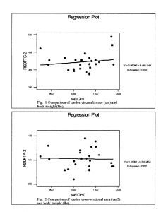
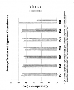
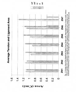
No significant difference was found between measurements of the tendons or ligaments of left and right forelimbs, therefore these values were pooled for greater statistical confidence. There was no correlation found at any zone between the circumference of tendons or ligaments and the weight of the horse (Fig. 1). There was also no correlation found at any zone between the cross-sectional area of tendons or ligaments and the weight of the horse (Fig. 2). The mean for the circumference and area of each tendon and ligament at each zone is presented in figures 3 and 4, respectively.
CONCLUSIONS
The results obtained are similar to other studies conducted on Thoroughbred and Standardbred Horses in relation to the overall shape and correlation of the ligamentous and tendinous structures. However, smaller measurements in both area and other measurements were apparent in this study, possibly due to the use of a smaller breed. The lack of correlation between indicated measurements and age was unexpected, but this may imply that tendon and ligament maturation, with respect to area and circumference are complete by two years of age. This offers an internal reference for comparison. The strong correlation between right and left legs in our study may be due to the symmetry of work these horses performed For those wanting to perform this procedure, it was also found when the alcohol had evaporated, quality monographic pictures were more difficult to produce. Therefore, application of more isopropyl alcohol is suggested before obtaining further measurements.
DISCUSSION
Ultrasound has become a useful diagnostic tool that provides quick and noninvasive viewing of soft tissue structures and facilitates differentiation and delineation of abnormal and normal size or echogenicityl. The results of this research project should provide equine practitioners with adequate data concerning normal tendonous and ligamentous metacarpal structures of the American Quarter Horse. Through utilizing these data, veterinarians can more effectively diagnose forelimb injuries, such as tendon and ligament strains or tears. Practitioners can also more objectively evaluate the progress of an injured Quarter horse’s treatment by comparing the measurements of the injured leg to that of the normal leg.
REFERENCES
- Hills A. Cary: Comparative Ultrasonic study of Normal Tendinous and Ligamentous Structures of the Palmar Metacarpus of Standardbred and Thoroughbred Horses. AAEP Proceedings 42:272-274, 1996.
- Smith RDW, Jones R, Webbon,PM: The cross-sectional areas of normal equine digital flexor tendons determined ultrasonographically. Equine Vet J 26:460-465,1994.
- Sande Ronald D, Tucker Russell L, Johnson Gary R: Diaanostic Ultrasound: Apilications in the Eauine Limb Eauine Diaanostic Ultrasonoaraphy Williams and Wilkins, Baltimore 103-117, 1998.
- Cartee Robert E etal: The Integument, Muscles, Tendons, Joints and Bones. Practical Veterinary Ultrasound Williams and Wilkins, Baltimore 268-271, 1995.
a. Pie Data 4800, Classic Medical Supply, Tequesta, Fla. 33649
b. Classical Medical Supply, Tequesta, Fla,33649


