Trisha L. Anderson and Dr. Noel L. Owen, Chemistry and Biochemistry
This project focused on endophytic fungi, fungi that live inside of plants without causing disease, as a source of compounds that may be useful in treating human disease. Endophytic fungi were chosen both because their relationship with the plants in which they live indicates that they provide some benefit to the plant and because they have a more specific relationship with a plant host, making acquisition of future samples of the same fungus more feasible. In addition, these fungal species have been less studied than many other aspects of natural products research. This research centered on the isolation of novel compounds that showed anticancer and antifungal activity.
Several plants were obtained from the San Rafael Swell and Cedar Breaks areas in Utah. Both of these sites were in areas that were relatively isolated, increasing the likelihood of finding new fungal species. Plant samples were sterilized and cut, and samples placed onto various media to facilitate fungal growth. After a total growth period of approximately 21 days, the fungi were transferred onto a liquid medium for another growth period of 7 to 10 days.
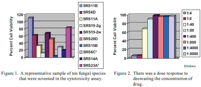
The liquid medium was dried, a solid sample weighed, and enough phosphate buffered saline, a solution which keeps the pH stable, was added to obtain final concentrations of 100, 50, and 25 mg per mL. Although these were not actual concentrations, since the fraction of the solid that was due to the compound of interest was not known, this procedure gave a rough estimate to use as a guideline in determining cytotoxicity. Screening for compounds that demonstrated anticancer activity was performed using an assay that employed HeLa cells, a type of cancer cell that has often been used in such research. Spectrophotometric analysis indicated the percent of cells that were still viable after incubation in the presence of the fungal product. An unexpected result of the drying and weighing procedure, however, was that none of our samples demonstrated anticancer activity, which prompted a reevaluation of the sample concentrations. After considering possible causes and performing further tests, it was determined that the most likely cause of the lack of activity was that the concentration was too low. As a result of this outcome, screening the samples straight from the liquid medium was begun. This procedure had several desirable outcomes. First, it allowed the screening process to occur much faster, as the samples did not have to undergo the drying process. Second, significant activity was evident in several of the samples. Twenty percent cell viability was chosen as our cutoff for determining activity of the samples. Although this level was chosen somewhat arbitrarily, it allowed us to keep our working samples at a manageable level while eliminating samples which showed less activity and hence less promise for further investigation. Differences in anticancer activity of several samples are clearly evident (Fig. 1). We screened 58 fungal species and found 20 to display some activity, a reasonable percentage with which to work. A dosage response is also seen when the concentration of the liquid medium is the assay is altered (Fig. 2).
An active sample in this assay, CB7C, was chosen to begin the extraction procedure. This fungus was grown in bulk, tested to ensure that it still displayed biological activity, and then extracted using various organic solvents. The solvent extraction that demonstrated the highest level of biological activity was the ethyl acetate extraction. A 2 mg/mL solution of the extract resulted in only a 1 percent cell viability. The extract was successfully dried and found to be active at much lower concentrations than in the original sample of liquid medium, at around 0.5 mg/mL.
Screening for antifungal activity was also performed. Some fungi produce chemicals, called antimycotics, which are not harmful to themselves but do inhibit the growth of other fungi. Screening for this activity involved placing a rapidly growing fungus, Pythium or Sclerotinia, in wells containing the liquid medium sample and relied on visual inspection to determine inhibition of fungal growth. Even with this cruder assay, some fungi showed evidence of significant angifungal activity. In the course of this research, however, it was hypothesized that because these chemicals are produced to deter competing fungi, perhaps some fungi only produce these antimycotics when there are other fungi present, thereby conserving valuable biological resources. Indeed, when we began growing three to four fungal strains in the same flask, the percentage of fungal species that displayed positive tests in the antifungal assays increased.
This work is ongoing. There still remains to further isolate and characterize these samples, as well as begin the extractions of other samples that have demonstrated activity. We will choose three compounds that have a high level of biological activity on which to do structural work. In addition, we will continue with the screening process to increase the number of potential candidates as some compounds will undoubtedly already be known and other will be unsuitable for medicinal use. These preliminary results have helped us to refine our screening process in order to better identify compounds that show promise in eventual medicinal use. Research on these endophytic fungi demonstrates that it appears to remain a promising area for continued investigation.