Jason Neff and Dr. Richard Vanfleet, Department of Physics and Astronomy
The original title submitted for my research was “Stacking Faults in GaN”. This study was aimed at employing the use of the Transmission Electron Microscope (TEM) to understand and image defects in the crystal lattice of Gallium Nitride (GaN) that lead to stacking faults. Because of difficulties in the preparation of the material we decided to use the resources and time granted us to work on a project in conjunction with some individuals in the chemistry department. This new project is consistent with the prior investigation, in that the same procedures would be followed, with the same equipment, but with different materials. Also in the old project our study was designed to characterize specific properties of GaN, and with the data collected in this assignment we would also be able to do that with Titanium Dioxide (TiO2) as well as label areas of interest for our collaborative group.
To begin research on a TEM one must be able to operate and understand how the components of this powerful instrument work. This is where my research began. I was enrolled in a 16 hour course that teaches the basics of a TEM, on how to run and operate these particular instruments. Because of the sensitivity of TEM’s, we were instructed on the functions of all major components and were also required to spend two sessions on the microscope with a trained microscopist. From there we were allowed to run the microscopes on our own with the aid of Dr. Vanfleet and Dr. Jeffery Farrer (Facilities Manager) close by.
A majority of my research was conducted on a 300 KV field emission TECNAI-F30-Super-twin TEM. This microscope is located in U183 of the Underground Lab in the Eyring Science Center.
After learning how to operate the TEM my responsibility was to acquire the samples and learn how to prepare them so we could study them in the microscope. We received two different phases of TiO2 (Anatase and Rutile) 1 nanoparticles in addition to their bulk particles, making in total four different powders to characterize. These samples were supplied by Dr. Boerio-Goates in the chemistry with hopes of understanding different behaviors expressed by components with the same chemical composition but with different surface/volume ratio. After preparing the sample our goal was to use the TEM to characterize these two phases which include its microstructure (grain size and orientation), particle size and shape, surface area, and density.
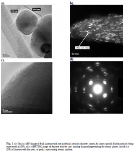
Using Bright and Dark Field imaging (BF&DF), Diffraction Patterns (DP) and High Resolution Images (HRTEM) for both phases, we were able to characterize the phase of Anatase nanoparticles to be in the range of 7-10 nm but to be non-distinct in larger agglomerates and polycrystalline. The same phase bulk particle is found to be of all sizes in the micron range and is a single crystal. We recognize the nanoparticle for Rutile to be rod like in shape with an
1 Guangshe Li, Liping Li, Juliana Boerio-Goates, and Brian Woodfield, “High Purity Anatase TiO2 Nanocrystals: Near Room Temperature Synthesis, Grain Growth Kinetics, and Surface Hydration Chemistry,”.
overall area of 7-20 nm and found to be distinct in larger agglomerates and single crystalline. Rutile’s bulk particle is more spherical, in the micron range, and single crystal. This information along with the images assisted Dr. Boerio-Goates and collogues in presenting their work in a few conferences and presentations. Follow up work with the transition of TiC to TiO2 is being planned.

