Ryan Earp and Dr. Steven Graves, Chemistry and Biochemistry
Nearly 20.5 million Americans above the age of 40 suffer impaired vision caused by cataracts. By the age of 80, the percentage of Americans suffering from cataracts increases to 50%. According to the World Health Organization cataracts are the leading cause of blindness in the world. The United States government spends an estimated $3.4 billion annually to treat cataracts surgically. The enormous cost of cataract treatment suggests a great need for non-invasive cataract treatment.
A number of studies have suggested a link between the formation of cataracts and a decrease in Na,K-ATPase (sodium pump or SP) activity. Research has demonstrated that the sodium pump located in the cell membrane acts to transport 3 sodium ions out of and 2 potassium ions into the cell. Loss of the SP activity would bring about sodium accumulation inside lens cells causing protein to become insoluble, precipitate and cloud the lens. Preliminary research conducted in our lab has shown that in addition to the SP present in the lens cell membrane, there are also SP units present in the cytosol of cells. Some researchers have suggested that factors present in the aqueous humor of the eye interact with the SP in the membrane of lens epithelial cells to reduce their activity, thus initiating or mediating cataract formation. Indeed, they have shown that aqueous humor acts to inhibit isolated Na,K-ATPase. However, preliminary research in our lab has shown that in response to SP inhibition, caused by ouabain (a plant derived specific inhibitor of the SP), there is a redistribution of the SP from the cytosol to the cell membrane. The critical issue is whether the increase in SP units in the cell membrane in response to this external inhibitor compensates for the inhibition and SP activity is maintained at a constant level. If SP activity remains constant, then another mechanism must be involved in the observed SP reduction in cataracts. If the net SP activity is less, despite movement of SP units from the cytosol to outer membrane, then this mechanism could explain part of the pathogenesis of cataracts.
The purpose of my project was to discover how ouabain affects the SP activity of bovine lens epithelial cells. Because SP units take extracellular rubidium (Rb) into the cell in the absence of potassium, I planned to use an Rb uptake assay to compare the activity of control cells to that of 24-hour ouabain treated cells. Then, using an atomic absorption graphite furnace, I would quantify the Rb inside of cells after washing them in an Rb solution for thirty minutes.
The data I obtained appeared to show that ouabain-treated cells had a lower concentration of Rb in their cytosol than control cells. This would suggest that in spite of mechanisms that appear to move SP from the cytosol to the membrane in response to ouabain inhibition, ouabain inhibited cells still exhibit less SP activity than control cells. However, due to significant and unresolved problems with our lab’s graphite furnace, the accuracy of the data I obtained is questionable. The amount of data I obtained is limited and inconsistent. Thus the results obtained are not statistically significant.
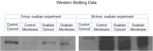
In addition to running the Rb uptake assay in order to determine SP activity, I also used Western Blotting Analyses to confirm earlier studies performed in my lab that suggest that SP units migrate from the cytosol to the membrane when lens cells are treated with ouabain. During this process I made an interesting discovery: when cells are exposed to ouabain for 24 hours the number of SP units in the cytosol decreases while the number in the membrane increases. This is consistent with previous experiments. However, when the cells were treated with Ouabain for only 5 hours the opposite occurred: the number of SP units in the membrane decreased while the number of units in the cytosol increased. I hypothesize that in an attempt to maintain uniform SP activity, the cell removes SP from the membrane that have been rendered useless by ouabain inhibition. This would explain the decrease in membrane-bound SP units at 5 hours. The cell then transports fully functioning SP units from the cytosol to the membrane in an attempt to stabilize the SP activity and reestablish sodium and potassium levels that are necessary for the cell to function properly. Because of the difficulties I encountered with the graphite furnace, I have not been able to show whether or not the cell is able to transport sufficient SP to the membrane in order to compensate for ouabain-inhibition.
I plan to continue investigating the problem by either using a different graphite furnace, or by making repairs to the furnace in our lab. In light of the discovery made regarding 5-hour and 24-hour ouabain exposure, I will analyze SP activity at both time intervals in order to gain a better understanding of the effects of ouabain inhibition of bovine lens epithelial cells.

These are photographic films of SP units obtained through western blotting electrophoresis. The first film shows a decrease in the number of SP units in the cytosol and an increase in the number membrane-bound SP units after 5 hours of exposure to ouabain. The second film shows a decrease in cytosolic SP and an increase in membrane SP after exposing lens cells to ouabain for 24 hours.
