Ghassan Sanber and Dr. Aaron Hawkins, Electrical and Computer Engineering
The fabrication and testing of micro-scale electroosmotic pumps, made with a thin-film deposition process compatible with standard silicon processing, is described. The fabrication method relies on chemical vapor deposition and chemical sacrificial etching to form long hollow fluid microchannels. Each pump consists of parallel narrow microchannels that deliver fluid to a larger wide sample handling microchannel. Flow rates were measured on these devices for different applied voltages using charge-coupled device (CCD) imaging. Pressures up to 1000 psi were produced. The pumps are based on the multiple open-channel electroosmotic pumping system described by Lazar et al [1]. However, instead of etching a glass substrate and using bonding to fabricate the pumps, thin film-deposition was used. This method of fabricating pumps is attractive because it can be used on a variety of substrates and offers high-scale integration with other planar thin-film components. These pumps have significantly smaller core diameters and spacing between microchannels than pumps made with alternative fabrication methods.
Fabrication Process
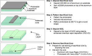
To build the microchannels a sacrificial core is patterned on the substrate and then coated with a thin film of SiO2. The core is then etched out leaving a hollow microchannel. The fabrication method described below uses photoresist on top of a thin aluminum layer as the sacrificial core. However, a variety of sacrificial materials can be used to produce different core geometries.

Electroosmotic Micropumping Principle and Pump Structure
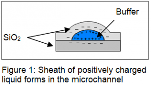
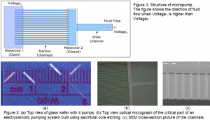
The outer oxide surface is negatively charged. This negative charge attracts positive ions in the solution forming a sheath of positively charged liquid around uncharged liquid. Applying a voltage differential across the microchannel creates an electric field. This electric field causes the positively charged sheath to move towards the negative electrode pulling the uncharged liquid core along and thus providing the pumping action. Several parallel microchannels share one reservoir and deliver pressure into one wider channel as shown in Figure 2. Figure 3 shows pictures of the pumps.


Testing the Micropump
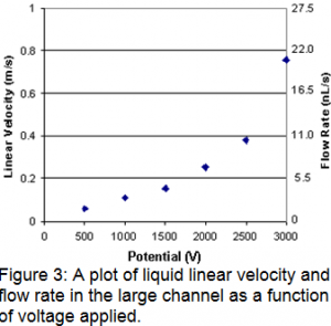
The pump was initially filled with a carbonate buffer solution. The reservoir surrounding the small channels was filled with carbonate buffer containing rhodamine B. Voltage was applied to the pump reservoir, and the pooled buffer at the opening of the large channel was grounded, driving the electroosmotic flow toward ground. The movement of rhodamine B through the large channel was followed using a CCD to image the laser induced fluorescence signal from the compound. Flow rates were determined by the time span between the initial appearance of rhodamine B and complete filling of the imaged channel.

References
- Iulia M. Lazar and Barry L. Karger, “Multiple Open-Channel Electroosmotic Pumping System for Microfluidic Sample Handling”, Analytical Chemistry, Vol. 74, No. 24, (2002).
