Kameron Hansen and Dr. John Colton, Department of Physics and Astronomy
Ferritin (FTN) is a spherical protein shell used nearly ubiquitously across life to store and transport iron in a non-toxic form. Ferritin’s natural occurring ferrihydrite mineral (Fe(O)OH) can be removed, leaving behind a hollow interior that conveniently serves as a template for researchers to grow quantum dots that are both spherical and confined to the protein’s inner diameter of 8 nm. The resulting protein-quantum dot constructs have been used as biological markers, photocatalysts, and photovoltaic photonabsorbers. In this study, we synthesize lead selenide and lead telluride quantum dots (QDs) inside ferritin (termed PbSe-FTN and PbTe-FTN, respectively) to make a material that has absorption and emission properties dominated by the PbSe/PbTe core and chemical properties dominated by the protein exterior. These chemical properties of ferritin have allowed the closely-related lead sulfide, in its ferritin-encapsulated quantum dot form (PbS-FTN), to be an effective inhibitor of cancerous cell growth while remaining non-toxic to other tissues. Since PbS, PbSe, and PbTe are closely related (all are direct, low-gap IV-VI semiconductors that share the same halite crystal structure), it is plausible PbSe-FTN and PbTe-FTN will also find application as cancer therapies. Alternatively, ferritin’s ability to be organized in structured arrays, it’s conductive shell, and its protection against photocorrosion are of relevance when considering the application of these materials to photovoltaics, which is an area PbSe QDs are commonly used due to their high probability for multiple exciton generation.
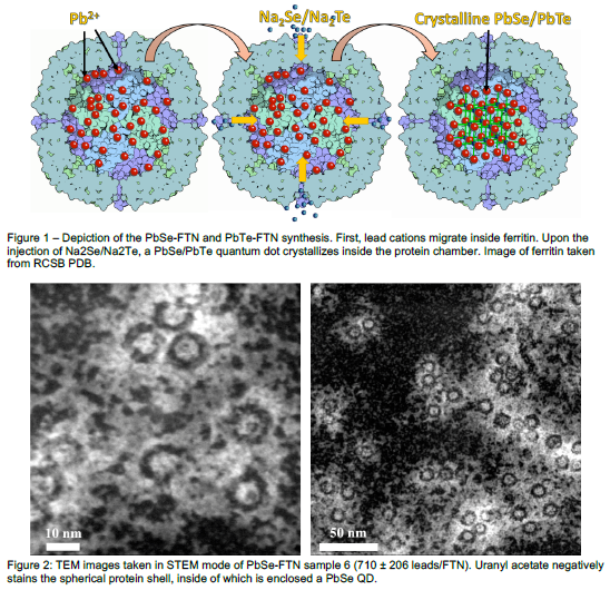
Core formation was achieved in PbSe samples 1 – 6 and PbTe samples 1 – 4, but not the additional samples that targeted more lead atoms per ferritin. Therefore, we recommend the synthesis parameters used for PbSe sample 6 and PbTe sample 4 if full core formation is desired; TEM images reveal these samples have core sizes of 7.03 (s = 0.79) nm and 7.32 (s = 0.68) nm while filling 91.8 % and 93.7 % of ferritin, respectively.
TEM images of PbSe-FTN (Figure 2) clearly reveal ~ 7 nm diameter cores inside the negatively-stained ferritin. Incomplete cores were observed to grow in a donut shape, with crystallization starting on ferritin’s inner surface. The presence of lead and selenium/tellurium inside ferritin was further corroborated by filtering samples through a size-exclusion matrix, harvesting the fractions with ferritin-sized solutes, and measuring the lead, selenium, and tellurium concentrations of these fractions. For PbSe-FTN sample 4, the protein, lead, and selenium concentrations were measured for each of the 15 fractions. The aligned peaks for all three concentrations demonstrate that these different-sized solutes fall through the size exclusion matrix at equal rates, i.e. the lead and selenium atoms are contained inside ferritin.
Our results show the new synthesis method for PbSe-FTN / PbTe-FTN successfully fills 91.8 % / 93.7 % of ferritin and can be used to make cores with an average diameter of 7.03 (s = 0.79) nm / 7.32 (s = 0.68) nm. These materials may be useful as cancer therapies or as photon absorbers in solar cells.

