
Devon Smith, Steven Cook
Tear Analysis: High-speed video capture of the Anterior Cruciate Ligament Tear
Dr. Jonathan Wisco, PdBio
Introduction
The tearing of the ACL is among one of the most common sports injuries seen during this era.
Research shows that annually there are more than 80,000 documented cases of ACL tears
occurring with a 15x greater possibility of re-tearing after reconstruction.1 Currently, there is no
digitally filmed data on how the fiber bundles unravel during an induced ACL tear. Published
studies utilize a Porcine ACL as the highest anatomically comparative model to Human ACL.
We are looking to create a grade 3 ACL tear in both human and porcine ACL’s, and film them at
high-speed while it unravels. We have created a stain made from powdered sugar and blue dye
that revealed both the posterolateral and anteromedial fiber bundle architecture. This allowed the
high-speed video camera to capture the ACL fiber bundles tearing from the femoral site in
greater detail. A deeper understanding of ACL tear microstructure, will aid in understanding why
current repair techniques are ineffective and how they can be improved.
Methodology
We filmed scalpel-induced tears of human and porcine anterior
cruciate ligaments (ACL) using a high-speed (6000 fps)
camera. The porcine ACL is routinely used as an ex vivo
animal comparison model of the human ACL. We dissected
and stained three opportunity unembalmed human specimens,
as well as three unembalmed porcine specimens to expose the
ACL, then stained the ACL’s with a blue dye solution we
developed, to provide high visual contrast of ligament
fibers. With the femur fixed securely on a table surface, and the
tibia hanging over the edge, resulting in a flexed knee joint at 90
degrees, a 2.2 kg weight was attached to the tibia in order to
induce tension on the ACL. The ACL was then cut anteriorly to a depth of approximately 1-3
mm with a scalpel, while a high speed video camera recorded the unraveling of the anteromedial
(AM) and posterolateral (PL) bundle fibers.
Results
There were noticeable differences in the unraveling of the human fiber bundles to the porcine
model. Human ACL fiber bundles were tightly oriented together in the knee, that there was not
any visible physical separation of the AM and PL fiber bundles. The AM and PL fiber bundles
were very similar in lengths, and it was relatively easy to put stress on either fiber bundle. Every
video tear showed a small ripple going through the AM fiber immediately prior to tearing. In all
three tears, the AM fiber bundle tore in an untwisting manner while the PL bundle acted as a post
for the AM bundle. The PL fiber bundle then tore in a linear fashion. During the tearing process,
both bundles tore in unison, one right after the other as if they were connected together.
Figure 1
Human ACL at the initiation of a tear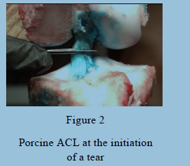
The porcine model displayed key features that differed relative to the human ACL model. First, the porcine ACL was comprised of two fiber bundles that were separate from each other by a 1-2 mm gap, and the geometry of the fibers were oriented in a triangular shape. In addition to this separation of the two bundles, it was apparent that the porcine PL bundle was shorter than the AM bundle. The final key difference was, due to their separation, the two bundles tore independently of each other. When the AM bundle was induced with a cut it would essentially cease to tear, until the PL bundle tore first.
Discussion
Utilizing a high speed camera to film the unravelling of the AM and PL bundles as the ACL tore
provided insights to their structural arrangement. Knowing the native structure of the ACL and
how the fiber bundles unravel is key to possibly reverse engineering better grafts and developing
more effective repair and reconstruction methods. Of particular interest in the human AM bundle
tears, was the ripple of a subpopulation of fibers after the tear was induced. It is likely that the
pressure induced by the cut, before it tears the fibers, places a subpopulation of fibers under
increased tension such that after they are cut, they release their energy as a ripple.
The porcine model demonstrated key differences to the human model that may provide valuable
insight to new methods by which the ACL could be reconstructed after it has undergone a tear.
Specifically, the separation of the two fiber bundles resulted in the bundles tearing independently
of each other. This may lead to increase stability within the porcine knee because the bundles
are not dependent on the stability of the other. Additionally, the AM and PL fiber bundles are
significantly separated at their insertion points, forming a triangular insertion into the tibia. We
surmise that this results in stronger porcine ACL structure, since the greater surface area of the
fiber bundles might lead to increase strength and resistance to tear.
Conclusion
This project focused on the filming of ACL tearing using a high speed camera. We were able to
visualize the unraveling of the ACL fiber bundles. We recognize that tensile and shear forces
were not measured; future work will combine videography with traditional biomechanics
methodologies. The knee was placed in a flexed position for convenience of its filming, but we
acknowledge that 90 degrees of flexion is not typically when the ACL tears. We would also like
to attempt filming while the knee is extended (or at least only slightly flexed). Furthermore, we
will explore twisting the femur and tibia in opposite directions, pulling the proximal tibial head
anteriorly, and placing a valgus force on the knee while in flexion; all stresses that the knee
experiences when the ACL is torn. It is our desire that these added measures will enable a more
accurate representation of the ACL within the motion of a natural tear without the introduction of
a cut.
References
1 – Griffin L, Agel J, Albohm M, Arendt E. Noncontact anterior cruciate ligament injuries: risk
factors and prevention strategies. J Am Acad Orthop Surg. 2000;8(3):141-50.