Kener, Kyle
Defining the role of Nr4a3 in β-cell function by analysis of full body knockout
Faculty Mentor: Jeffery Tessem; Nutrition, Dietetics, and Food Science
The prevalence of diabetes continues to grow at an alarming rate. Both type 1 (T1D) and type 2 diabetes (T2D) lead to an eventual destruction of β-cells and an inability to maintain a state of normoglycemia. We aim to increase functional β-cell mass which could potentially cure diabetes. The orphan nuclear receptor Nr4a3 is a key link in the Nkx6.1 proliferation pathway. It has been shown to play an important role in β-cell proliferation and glucose stimulated insulin secretion (1,2)
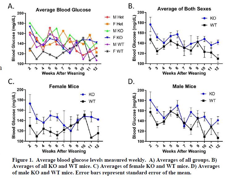
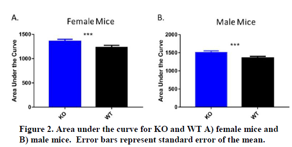
Using our full body Nr4a3 knockout (KO) mouse model, our first focus has been on insulin secretion. We have taken weekly blood glucose measurements (Figure 1). We compared the difference between mice homozygous for the Nr4a3 deletion (KO), heterozygous for the deletion (Het), and wild type (WT). Although blood glucose levels of the mice demonstrate different levels of variation, on average Nr4a3 KO mice have higher blood glucose levels than the WT mice for both males and females. When analyzed as area under the curve  (AUC), Nr4a3 KO mice are significantly greater in both sexes (Figure 2). This could
  demonstrate that the Nr4a3 deficient mice have an impairment in β-cell function, specifically insulin secretion, or in insulin signaling in peripheral tissue. A reduction in ability to secrete insulin would lead to higher blood glucose levels. This is what happens in T1D due to β-cell apoptosis and T2D due to beta cell dysfunction. Some groups from this data set have more mice than others with group sizes ranging from 2 to 7 mice. We will continue to update this data set with more mice from each group as we get them. Our goal is to have data from 10 mice for each group.
To further analyze insulin secretion, we have also administered oral glucose tolerance tests (GTT) at 1, 2, and 3 months after weaning (7 weeks, 11 weeks, and 15 weeks after birth). To perform GTTs, mice are fasted for a minimum of 12 hours prior to injection. Mice are injected in the peritoneal cavity with a 20% glucose solution adjusted for their weight. For example, a 20.0-gram mouse would be injected with 0.2mL of solution and a 25.0-gram mouse with 0.25mL. These test have resulted in a broad range of responses and all groups seem to follow a similar trend of glucose clearance. We expected to see a reduction of insulin secretion in KO mice, which would lead to high blood glucose levels for an extended period of time when compared with WT mice. However, as observed in Figure 3, the KO mice seem to be clearing glucose from the blood stream at about the same rate as the WT mice. The 3 month GTT has the fewest number of mice represented which is a likely cause of the current variation.

Analysis of β-cell area and replication by means of staining fixed pancreata for insulin, Ki-67, yH2AX, and PHH3 is also underway. Pancreata, along with other organs, have been harvested for this experiment. Fixation and staining will begin within the next couple months.
Preliminary data from these experiments were presented at the Utah Conference of Undergraduate Research in February 2016. Final data from these experiments will be analyzed and submitted for publication in a peer-reviewed journal. We anticipate publication of these results in 2018.
Sources
1. Tessem, J.S. et al., Nkx6.1 regulates islet β-cell proliferation via Nr4a1 and Nr4a3 nuclear receptors. Proceedings of the National Academy of Sciences. 111(14): 5242-5247. (2014)
2. Weyrich P, Staiger H, Stancakova A, et al., Common polymorphisms within the NR4A3 locus, encoding the orphan nuclear receptor Nor-1, are associated with enhanced β-cell function in non-diabetic subjects. BMC Medical Genetics. 10:77 doi:10.1186/1471-2350-10-77. (2009)
