Seth Spencer and C. Brock Kirwan, Psychology
Introduction
Many studies have shown a correlation between physical exercise and healthy cognitive processing. It has been shown extensively that regular exercise has a positive impact on brain health. One example is a study that linked increased exercise with greater performance on as shown via a Stroop Color-Word test [1]. Similarly, regular exercise has been shown to be related to improved memory and object recognition [2]. Another study has shown that even a single session of exercise can alter functional connectivity in the brain as measured by the resting-state fMRI [3]. The problem with this literature is an utter lack of universal standards that constitute exercise. Most studies split a group of individuals into “high” and “low” exercisers. Sometimes the “high” exercisers are Olympic athletes—an impractical standard of exercise for the general public.
In this study we will use the standards of exercise set forth by the American Heart Association to provide a common convention for a scattered field of literature. Indeed, to our knowledge, there have been no studies that examine specifically how meeting the physical activity guidelines prescribed by the American Heart Association (AHA) impact cognitive function. Our hypothesis is that individuals who engage in high exercise (meet AHA recommended levels of physical activity) will have greater pattern separation (PS) scores, activation of CA3/DG, and increased white matter integrity in cingulum, uncinate fasciculus, and temporal lobe pathways, which have been established for their connection with memory [4]. A secondary goal of this experiment was to explore whole brain activation associated with pattern separation.
Methods
For this study we recruited 49 individuals (22 males, 27 females median age = 24 years). Participants were divided into three groups of high exercise (met the levels of the American Heart Association; n = 18), light exercise (did not meet AHA recommendations; n = 14), and sedentary (no exercise; n = 17). The study was non-interventional, and exercise levels were established based off of results of a questionnaire and a movement meter (actigraph) which was worn for a week. Cognitive health was assessed through the means of a behavioral pattern separation task which was done concurrent with an fMRI scan. A DTI scan was performed at this same time.
Behaviorally, participants were required to distinguish between highly similar images. Each image presented was either a new stimulus (foil), a repeated stimulus (old), or an image which was similar, but not exactly the same as a previous image (lure). Their responses to these images were categorized as CR (foil image called new), Hit (repeat image identified as old), L-CR (lure image identified as similar), and L-FA (lure image identified as old).
An fMRI scan was performed during this behavioral task to assess sub-hippocampal activation.
Results
A one way ANOVA was performed on the participants responses which revealed no significant differences between groups by response on the PS task. There were also limited differences found between the exercise groups based on physical activity of the movement meter. Only the high and sedentary groups were significantly different.
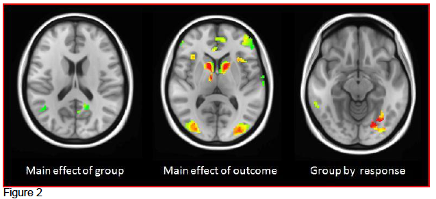
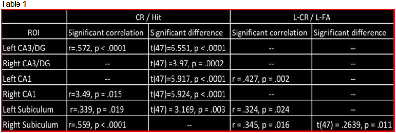
A repeated-measures ANOVA was performed to assess results of the fMRI scan. The regions of interest (figure 1) were the CA1, CA3/DG, and subiculum. There was no significant differences between the groups, but there was a significant difference seen in hippocampal activation based on response (CR, Hit, L-CR, L-FA; see table 1). Whole brain analysis also revealed 3 functional areas by main effect of group, 21 functional areas by main effect of response, and 5 functional areas of group by response (see figure 2).
MANOVAs were performed on the DTI scans which revealed significant group differences in bilateral cingulum, left middle temporal gyrus, and right uncinate fasciculus (see figure 3).
Conclusions
Although groups did not differ by PS score (behavioral) or ROI (fMRI), as expected across groups, there was a significant interaction found between the response outcomes and hippocampal subregions. The group differences by whole brain analysis maybe a result of those areas involved in concentration, or cognitive function associated with our particular memory task. Similarly, the DTI results need to be further explored to differentiate their role in memory, or this task specifically. Additional studies which use an implicit version of this task may also be helpful in the future.
Scholarly Sources
- Vasques PE, Moraes H, Silveira H, Deslandes AC, Laks, J. (2011) Acute exercise improves cognition in the depressed elderly: the effect of dual-task Clinics. Clinical Science [Sao Paulo] 66(9):1553–1557.
- Hopkins ME, Davis FC, VanTieghem MR, Whalen PJ, and Bucci DJ. (2012) Differential Effects of Acute and Regular Physical Exercise on Cognition and Affect. PMCID: PMC3374855 Neuroscience. 215: 59–68.
- Rajab AS, Crane DE, Middleton LE, Robertson AD, Hampson M, MacIntosh BJ. (2014) A single session of exercise increases connectivity in sensorimotor-related brain networks: a resting-state fMRI study in young healthy adults. Frontiers in Human Neuroscience, v.8: 625. PMC4132485
- Bakker, A., Kirwan, C. B., Miller, M., & Stark, C. E. (2008). Pattern separation in the human hippocampal CA3 and dentate gyrus. Science, 319(5870), 1640-1642. doi: 10.1126/science.1152882