
Jack Silcox and Dr. Bruce Brown, Psychology Department
Introduction
Many have recognized the importance of identifying biomarkers for cognitive disorders (Casey, Craddock, Cuthbert, Hyman, Lee and Ressler, 2013). This is due to the amount of subjectivity that exists in current diagnostic methods. A patient with a mental disorder has to rely upon a psychiatrist’s experience and limited understanding of the patient’s mental state for a diagnosis, with the hopes of receiving the most effective treatment. Finding biomarkers to help supplement a psychiatrist’s subjective diagnosis would allow a patient to receive the appropriate treatment at the appropriate time. Current brain imaging technology has the potential for identifying biomarkers for mental illnesses and has already helped make tremendous advances in understanding mental illness.
Of the brain imaging technologies available, the electroencephalogram (EEG) is the least expensive and least intrusive (Luck, 2005). EEGs capture an incredible amount of detail about a person’s current mental state. Unfortunately, current methods of analyzing these data have not proven successful as diagnostic tools. This study looked at developing and refining a new method of analyzing data collected from an EEG. In this study we attempted to use our method to distinguish between male and female differences while performing a cognitive task. This seemed like an appropriate way to test the power of this method in finding differences between groups of people with different mental conditions.
Methodology
We used data collected by Dr. Bruce Brown and Dr. Dawson Hedges back in 2003 (Brown, Hedges and Gant, 2008). Data was collected on 51 subjects wearing an EEG with 5 locations (Fz, Oz, Cz, T3, T4). The study was designed after the classic study done by Jonides and Gleitman (1972) in which subjects had to detect letters or numbers against a field of letters or numbers. The advantage of using this old data set is that we will be able to compare the results we find using our new method against the results obtained from the method used by Brown, et. al originally.
Most methods of analyzing event-related potentials (ERP) use an ANOVA test on small portions of an entire contour averaged for all subjects (Luck, 2005). However, this does not allow one to test the effects of experimental manipulations holistically. When Brown, et. al(2008) originally ran analyses of their data they used an extraction of principal components from the entire event-related potential contour as a group analysis of all the subjects combined. This yielded fairly good results. We hypothesized that if principal component analysis is done on one subject and one electrode location at a time for the full ERP contour and then we regress the principal components, we will be able to extract cognitive components and we will obtain higher Fratios.
In other words, we felt that we should be able to get the measurement error out of the analysis when we look at the data for one person and one location at a time. These regressed principal components will act like “finger prints”, or personal identifiers, for each individual person. We hypothesize that we can use these “finger prints” to discriminate between genders when we run a similar analysis on each person at each electrode location.
Results
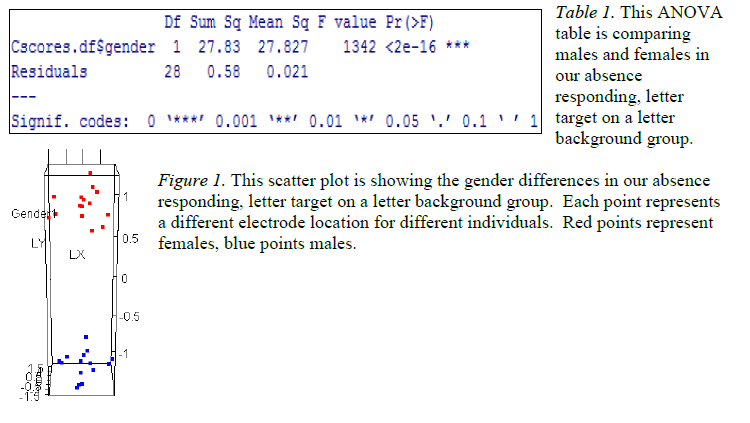
The EEG data that was analyzed with our methodology consistently showed differences between males and females while performing the cognitive task (P<0.0001). Table 1 is representative of 24 different comparisons that were done. Figure 1 visually displays that there was no overlap between genders while performing the task, which was consistent for all 24 comparisons.
Discussion and Conclusion
Recent studies have found gender differences in brain structure (Verma, 2014). However, we found that not only are there structural differences but there are also functional differences in male and female brains as analyzed by EEG.
Our new methodology was surprisingly sensitive to gender differences while performing the cognitive task. The F-ratios we found were on average about 100 times larger than those found with the traditional method. This suggests that this new method has incredible potential for use in diagnostics. There is a need for more refinement of the method and the technology that uses it but that is currently underway.
Scholarly Sources
Brown, B. L., Hendrix, S. B., Hedges, D. W., and Smith, T. S. (2012). Multivariate Analysis for the Biobehavioral and Social Sciences. New York: John Wiley & Sons, Inc.
Casey, B. J., Craddock, N., Cuthbert, B. N., Hyman, S. E., Lee, F. S. and Ressler, K. J. (2013). DSM-5 and RDoC: progress in psychiatry research? Retrieved from http://www.nature.com/nrn/journal/v14/n11/abs/nrn3621.html
Luck, S. J. (2005). An Introduction to the Event-Related Potential Technique. A Bradford Book; 1 editionJonides, J., and Gleitman, H. (1972). A conceptual category effect in visual search: O as letter or as digit. Perception and Psychophysics, 12, 457-460
Brown, B. L., Hedges, D. W., and Gantt, E. E. (2008). Brain Processes and Holistic Isomorphism: Moving Toward a Humanistic Neuroscience. Journal of Theoretical and Philosophical Psy. Vol. 28, No. 2, 2008, 356-374.
Ingalhalikar, M., Smith, A., Parker, D., Satterthwaite, T. D., Elliott, M. A., Ruparel, K., . . . Verma, R. (2014). Sex differences in the structural connectome of the human brain. Proceedings of the National Academy of Sciences of the United States of America, 111(2), 823-828. doi:10.1073/pnas.1316909110