Mariana L. F. Castroand Dr. David Johnson, Department of Anthropology
INTRODUCTION
The purpose of this project was to examine the paleopathologies of Nabataean burial remains, with emphasis on the osteological evidences for diet, sex and age. The main goal of Archaeology is to uncover the past. However, this cannot be done if scholars and students do not engage in the process of excavation, analysis and consequent publication of the results. This project was important insofar that it put forward a better understanding of Nabataean burial practices and their treatment of the dead. This study can also help us better understand human limitations and past socio-cultural dilemmas, including gender related issues, political hierarchy and dietary preferences.
The project described in this report consists of the methodical analysis of human bone remains from the Nabatean tomb 676, in Wadi Mataha (Site 15), Petra, Jordan. Its prevailing and meticulous façade points to the importance and possible royalty status of the people buried there. So far, all the remains found in the six loculi excavated (bed-rock cuts where the body of the deceased would be deposited) belong to seven women and one child, some with clear signs of deadly paleopathologies (Johnson, Ure, and Castro 2014). In the course of 6 weeks between May and mid-June of 2014, I oversaw an excavation in Tomb 676. During that time, we excavated two cists (L12 and L14), and recorded and examined all the human bones, artifacts, and stone. We discovered the remains of three people, yet only one of the them had evident signs of paleopathology.
METHODOLOGY
The methods for successful Osteology analysis include the recording of age, sex, and ethnicity (if possible). Furthermore, there must be a list and description of all the bones recovered from the burials, and a careful examination of each in order to determine any sign of paleopathology (trauma, inflammatory, circulatory, metabolic, neuroplastic or neuromechanical).
Bones
In order to identify each bone, I used Tim White’s Human Osteology (2012). This is the best reference for osteological analysis accessible to students of archaeology, and it was compiled by anatomists, anthropologists, and doctors. I used the basic anatomical terminology to identify the bones.
Sex
Sex and Gender should not be confused. Gender is an “aspect of a person’s social identity” (White 2012:408), whereas sex refers only to biological characteristics. There was nothing in either tomb that directly indicated gender. It is also important to note that the scientific determination of sex is surrounded by uncertainty. Archaeologists are constantly forced to used the terms “probable” and “possibly,” and the truth is that we can only be somewhat dependent on our confidence. In any case, there are some osteological clues that can help the determination of sex. These are the most helpful:
- The size and robustness of the Mastoid Process (Temporal Bone). The more prominent and robust the mastoid process is, the more likely it is a male.
- The prominence and sharpness of the Supraorbital Ridge (Skull). The sharpest it is, the more likely the individual was female.
- Smoothness and projection of the Mental Eminence (Mandibule). The more smooth and the less projected, the more likely it is a female.
- Probably the best indication of sex, the size of the Greater Sciatic Notch (Pelvis). The wider and bigger it is, the more likely the individual is female.
Age
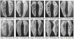
Unless there is a stele on the grave that indicates the time of birth and death, it is virtually impossible to determine someone’s exact age only through their bone remains. Because of this, in archaeology, age is always given in intervals, long or short, depending on how much the skeleton had matured at the time of death. It is easier to determine the exact age of younger children because bones tend to merge at specific times in someone’s life. After the growth stage, age is mostly determined by the deterioration of the friction points in the body (teeth, joints, etc.). The best indicators of age are:
- Teeth: teeth formation starts in the embryo at 14-16 weeks. Between 0-18 years, there are specific patterns of teeth growth. Nevertheless, after 18 years, age can only be accessed from teeth through ware. There are four distinct phases of emergence of the human dentition between 0-18 (White 2012:385):
- Most deciduous teeth emerge during the second year of life;
- The two permanent incisors and the first permanent molar usually emerge between 6 and 8 years;
- Most permanent canines, premolars, and second molars emerge between 10-12 years.
- The third molar (wisdom teeth) emerges around 18 years.
- Cranial suture closure: cranial bones fuse progressively as the individual ages. The shenoocipital synchondrosis fuses between 20-23 years old, and is the best marker of age in the Occipital bone.
- Bone length: not very reliable, but useful for relative dating.
- Pubic Symphyseal Surface: one of the most used and reliable indicators of age mainly because this is a part of the skeleton that continues to change as the body matures. More ridges and best shape indicates younger individuals
Disease
Disease refers to antemortem biological or environmental modifications of the skeleton. In particular, I will be discussing paleopathologies, which have been defined as the “science of the diseases which can be demonstrated in human and animal remains from ancient times (Ruffer 1913). Sometimes, trauma is included, since it is also considered pathological. However, in the scope of this report I will only discuss diseases that cause bone modification. Also, I will not discuss in detail each disorder and disease and their effects on bones, unless there are overarching characteristics seen in all manifestations of the disorder and the disease.
- Inflammatory/Immune: includes Leprosy, tuberculosis, Syphilis.
- Circulatory: affects the blood vessels, and it is evident on bones through the foramen, the marrow, and the surfaces where the muscles attach.
- Metabolic: includes dwarfism and gigantism, but also nutritional deficiencies. Generally we see an increase or a reduction in amount of bone tissue present. The bone affected is normally poorly mineralized.
- Neuromechanical: includes multiple sclerosis and osteoartiritis. Normally wear on the bones, and very related to the type of work activity. Visible in the femur, vertebrae, and feet and hand joints.
- Neoplastic: abnormal mass or tissue that is porus and darker, normally cancer.
RESULTS
Over the course of two months, our team excavated three individuals. There were two females in Cist 12 (Loculi 12), and one male in Cist 14 (Loculi 14). The women were probably disposed in the cist in a manner of secondary burial. In this type of enterrerment, after an individual dies he or she are left in the open to decompose, and then moved to another location where they are buried. The male, on the other hand, was an articulated burial (i.e., laid down on his back across the cist). See Annex 1 for more information on the osteological findings.
DISCUSSION AND CONCLUSION
There was no evidence of paleopathologies in Cist 12. This is not surprising, since we do not have many bones from the two females who were buried there. It is also strange that most of the bones from this cist were skull bones. From the teeth and mastoid processes, I was able to identify two probable females, a 8-12 years old and a young adult. It should also be noted that this cist was notorious for the artifacts that it revealed. We found 10 gold pieces of jewelry and nine beads made of carnelian and other rare stones (Figure 1).
Cist 14, the grave where the young 20-25 year old male was buried, revealed a different story. From the lesions of the calcaneous (Figure 2) and lumbar vertebrae (Figure 3), I concluded that this man was infected with skeletal tuberculosis (osteo-articular tuberculosis), and that he probably died from a combination of non-osteological symptoms that follow the bone evidence. Skeletal tuberculosis causes well defined lesions on the bone, similar to corrosion, and can affect “the spine, hip, knee, foot, elbow, wrist, hand, shoulder and as diaphysial foci.”1 In some cases, a multifocal skeletal TB can occur, which “is defined as osteoarticular lesions that occur simultaneously in two or more locations, with or without pulmonary involvement. It may affect a single bone with multifocal lytic cortical lesions or multiple sites at different bones.”2 The man’s left tibia displayed signs of cortical lesions (Figure 4).
Tuberculosis affects almost every organ in the body, but skeletal tuberculosis is always triggered by a primary lesion in the lung. According to Dr Yuranga Weerakkody and Dr Prashant Mudgal et al. , “The predisposing factors are protein energy malnutrition, environmental conditions and living standards such as poor sanitation, overcrowded housing and slum dwelling.”3 First century Petra was probably a place where these conditions thrived, and therefore skeletal tuberculosis is the best paleopathological fit for this man’s disease and likely cause of death.
1http://radiopaedia.org/articles/skeletal-tuberculosis
2http://www.ncbi.nlm.nih.gov/pmc/articles/PMC3524008/
3http://radiopaedia.org/articles/skeletal-tuberculosis
Annex 1
Osteological findings:
L12
- SU (Stratigraphic Unit) 3
- Rib fragment (3)
- Sphenoid, posterior fragment (1)
- Sphenoid, inferior fragment (1)
- Sphenoid, greater wing fragments (2)
- Mandibule fragment (1)
- Second incisor (deciduous)(fragment) – lateral, right, bottom.
- Unidentified fragments (14)
- SU4
- Parietal, left fragment (1)
- Occipital fragment (1)
- Frontal crest fragment (1)
- Orbital fragment, supraorbital margin (1)
- Sharp, Female
- Frontal fragments (3)
- Parietal unidentified fragments (7)
- Skull unidentified fragments (11)
- Maxilla, right side fragment (1)
- Teeth:
- Molar fragments (3)
- Teeth crown (2 fragments with roots)
- Pre-molar fragment (1)
- Teeth are not worn, no signs of cavities.
- Left temporal mastoid process (1)
- Small, Female
- Right eye socket fragment (1)
- Possible zygomatic fragment (1)
- Maxilla, lower eye socket fragment (1)
- Root fragments without caps (6)
- Possible palatine (lateral fragment) (1)
- Possible left conchal (1)
- Sphenoid, anterior, right, fragment (1)
- SU6
- Left parietal fragment (large) (1)
- Skull fragments (14)
- Frontal fragment (1)
- 1st metacarpal (right side) (1)
- 8-12 years old
- Distal phalanges (3)
- Alveolar process fragment (1)
- Mandible with teeth (uneupted molar )(1)
- 3rd molar without roots (unerupted?) (lower) (1)
- Deciduous mandibular molars (left and right) (2)
- Premolar fragment (1)
- Rdc1, Ldi2, Rdi2, Rdi2, Ldi2, di1
- 6 teeth: possible mandibular canines/incisors (4), premolars (2)
- All permanent teeth that have not formed roots yet
- 8-12 years old
- Cervical vertebra fragment (lower, 5 or 6), fragment (1)
- Rib fragments (25)
- Possible sternum fragment (1)
- Unidentified fragments (23)
- Cranial fragments (24)
- Rib fragments (5)
- Cervical vertebra (1 fragment)
- Right side temporal fragment with mastoid process (1)
- Unidentified fragments (various)
- Mandible pieces articulated (2)
L14 – Almost complete human skeleton of a male
- SU 5
- Dentition:
- Mandible:
- RI1, RI, RC1, RP1, RP2, RM1, RM2, RM3
- LI1, LI2, LC1, LP1, LP2, LM1, LM2, LM3
- Maxilla:
- RI1, RI2, RC1, RP1, RP2, RM1, RM2
- LI1, LI2, LC1, LP1, LP2, LM1, LM2, LM3 (not fully erupted)
- Notes:
- Mental eminence – p.364 – score 2/3
- No evident diseases on the bones or teeth (not chipped. Cracked, or transparent – very healthy teeth)
- All teeth erupted including M3’s – > 20 old
- No root transparency – < 40 old
- Based on ware of teeth – more on incisors – 20-30 old (more towards 20 years old).
- Arms
- Humerus:
- Left: 4 fragments, fractures post-mortum
- Right: 2 fragments
- Head 4.5 cm, whole bone 33.5 cm
- White stature 3.08 x 33.5 +70.45 = 173.63 cm
- Radius:
- Right: 4 fragments
- Left: 4 fragments
- Ulna:
- Right: 3 fragments (whole bone 24.3 cm)
- Left: 5 fragments
- Scapula:
- Fragments of both right and left
- Clavicle:
- Right: complete, 2 fragments
- Robust and shorter – probably right handed
- Left: not complete, 2 fragments (medial parts)
- Wrist bones:
- Left:
- Lunate
- Hamate
- Trapezium
- Trapezoid
- Capitate
- Triqueteral
- Right:
- Scaphoid
- Pisiform
- Trapezium
- Trapezoid
- Capitate
- Hand bones:
- Left (came from the left side of the loculus)
- Metacarpal: 1,4,5 + 1 shaft + 3 heads of metacarpals
- Proximal phalanges: 4,5
- Right (came from the right side of the loculus)
- Metacarpals: 3,4,5 (all broken on their proximal ends)
- Proximal phalanges: 5 +1 head
- Unidentified side:
- Intermediate phalanges 2,4,5 +1,2,3,4,5
- Distal phalanges 2,3,5 + 3
- Metacarpal 1
- Proximal phalanges 1,2,3,4 +1,2,3
- No perceivable difference in wrist bones and hand bones to determine handedness.
- Skull
- Frontal:
- 6 fragments including both supraorbital notches, broken frontal crest
- Parietals:
- Both left and right are complete
- Occipital:
- Broken into right and left fragments (complete). Internal occipital crest, complete foramen magnum.
- Temporal:
- Both temporal bones.
- Right mastoid process very protruding – probably male.
- Left temporal complete but missing the zigomatic process
- Zygomatic arches:
- 2: left and right. Right side is complete, and left is broken on the inferior medial end.
- Pelvis:
- Os coxae (both) – several fragments 8
- Symphysis: probably male in the Suchey-Brooks scoring system (White 2012:357) it score a 2, meaning the age range is limited to 19-34.
- Sciatich notch (Walker 1994) scoring system 3/2
- Sacrum: very fragmentary pieces
- Vertebrae:
- C4, c5, c7, c unknown (2x), T1-12 (8 are broken), L1-5 (4 and 5 present paleopathologies).
- Ribs:
- All 12 ribs – all medial endings of right ribs are present
- 11 medial endings on left side including 1st rib (2 pieces but complete)
- Sternum:
- Mostly compete manubrium
- Fragmented corpus sterni
- Legs:
- Femur:
- Right: 45.5 cm, 16 fragments including head and patellar surface
- Left: 46 cm, 12 fragments including head and patellar surface
- 170.89 cm (height from femur)
- Patella:
- Right and left intact
- Tibia:
- Left: 1 piece with both ends broken off
- Right : 2 pieces
- Fibula:
- Left: 3 fragments, missing proximal and distal ends
- Right: 2 fragments, including ends
- Feet:
- Left:
- Metatarsals: 1,2,3,4,5
- Proximal phalanges: 1,2,3,4,5
- Intermediate phalanges: 2,3,4
- Distal phalanges: 1,4
- Talsals:
- Cuneiform (medial)
- Right:
- Metatarsal: 1,2,3,4,5
- Proximal phalanges: 1,2,3,4,5
- Intermediate phalanges: 2,3,4
- Distal phalanges: 1
- Tarsals:
- Calcaneus (bone loss, sign of paleopathology)
- Talus
- Cuboid
- Navicular
- Cuneiform (medial)
- Intermediate cuneiform
Images
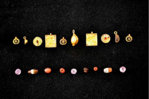 Figure 1. Gold Artifacts from Cist 12.
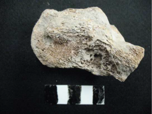 Figure 2. Calcaneous of male in Cist 14.
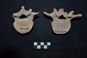 Figure 3. Lumbar vertebrae of man in Cist 14.
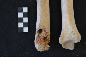 Figure 4. Tibia of man in Cist 14.
Scholarly Sources
2014: Johnson, David, Scott Ure and Mariana Castro. Interim report on four seasons of excavation of Wadi Mataha Site 15, BD Tomb 676. Annual of the Department of Antiquities of Jordan, Amman.
2000: White, T. D., and Pieter A. Folkens. Human Osteology. San Diego: Academic, Print.


