Dustin Day and Professor Alonzo Cook, Chemical Engineering
Introduction
A common treatment for a dysfunctional blood vessel due to atherosclerosis is the autologous transplant of a patient’s own veins. Removing portions of a patient’s vein to substitute for an artery often presents difficulties to surgeons because of the diseased state of the transplant vessels. However, the search for an ideal arterial substitute has recently taken exciting new turns and promises to bring new and efficient innovations to the field. The newest research focuses on reconstructing arteries from a patient’s own cells in vitro then implanting the lab- engineered tissue as a vascular substitute. Advantages to this method include a lack of adverse immunological responses from the graft recipient and ensuring that all the characteristics of living arteries are intact. Our blood vessel research team has entered the tissue engineering field in an exciting effort to 3D print cell-seeded vascular constructs.
Our initial goal is to print living endothelial and smooth muscle cells into the shape of human arteries. Using a variety of hydrogels as placement media, cells can be printed directly into shapes resembling multilayered artery tubules, stimulated to proliferate, and subsequently form functional arteries. When in contact with one another, smooth muscle and endothelial cells have been shown to secrete and reform the extracellular matrix of a vessel essentially recreating an authentic, naturally functioning human tissue. Multiple methods are also in place to assess the functionality of a vessel construct: tensile strength is measured, blood-tissue interactions are determined, and synchronization of smooth muscle contractions can be ensured. Finally, animal tests are performed to identify complete functionality in an organism and assess immunogenic reactions to the new tissue.
Methodology
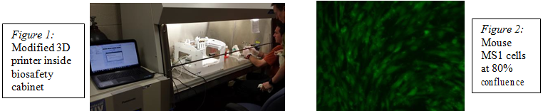
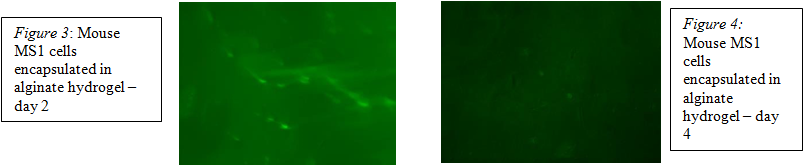
We have successfully assembled and modified a 3D printer to print living animal cells and we have encapsulated porcine aortic endothelial cells (PAEC) and MS1 mouse endothelial cells into a seaweed-based alginate hydrogel to facilitate the delivery of the proper cells to specific areas of the construct. Cells were cultured to 80% confluence (figure 2), pelleted, and homogenized with 10 mL of 1% alginate hydrogel that we previously prepared. The cell/gel mixture was then loaded into the 3D printer and various shapes were printed onto cell culture dishes. After printing we cross-linked the alginate with CaCl2 solution to solidify the gel matrix. Each of the printed shapes containing live cells were bathed in cell growth medium and monitored using fluorescent microscopy to track cell proliferation.
Results
Fluorescent images showed that the cells were able to survive the printing process and maintain viability in the alginate hydrogel, albeit with a brief lifespan. Figure 3 shows that cells were viable on day 2 after printing, but figure 4 shows no live cells indicating that there was minimal survival of cells past the third day after printing.
Discussion
The bright areas in figure 3 indicate living cells or clusters of cells within the alginate matrix showing signs of branching and attachment. This confirms that the endothelial cells indeed survived encapsulation and printing without being compromised. However, in the images taken after day 4 (figure 4) no live cells were detected. The absence of living cells four days after printing shows that there was a problem with cell replication and proliferation. Some possibilities could include the inability of the growth medium to reach the cells through the alginate matrix or the inability of the cells to properly attach to the hydrogel matrix and fill in space.
Conclusion
Our research demonstrates that we were able to successfully 3D print endothelial cells encapsulated in alginate gel. The branching that was seen is indicative of cell growth and attachment, but its disappearance after day 4 suggests that there could have been a problem with the attachment. In addition, if the cells were unable to reach the growth medium through the alginate gel matrix they would not receive the proper nutrients and would not survive replication. Therefore we conclude that 3D printed endothelial cells are able to survive encapsulation and printing, but their viability declines after day 2. The decline could be through an attachment mechanism malfunction or the inability of growth medium to reach the cells through the gel matrix. We are currently testing methods to increase porosity and modifying the alginate through sulfonation and methacrylation to make a more hospitable environment for the cells.