
Baba, Tomonori
Tissue Engineering: Cell-Seeded 3D Printed Vascular Graft
Faculty Mentor: Alonzo Cook, Chemical Engineering
Introduction
The purpose of this project was to create and test a biocompatible modified alginate gel for 3D printing blood vessels to be used in vascular networks for tissue engineering and as vascular grafts. Vascular disease is the leading cause of death in the United States, and more than $100 billion are spent annually for its treatment. Treatments for cardiovascular disease, such as synthetic blood vessels still have some problems with thrombosis and immune rejection. We propose to use the patient’s own cells to form replacement blood vessels, to reduce the risk of immune rejection and the endothelial cells will be able to create a naturally non-thrombogenic surface that can renew itself. Another treatment, endoscopic vessel harvesting, requires additional surgery to remove a section of blood vessel from one location and place it in the region of interest for treatment. This involves the danger of internal bleeding following the surgery. With our approach there will be no need for endoscopic vessel harvesting. This new bioprinting approach requires a gel with certain characteristics: biodegradable, maintain cells viable, crosslinkable, and 3D-printable. This project produced a gel that meets those requirements.
Methodology
Alginate (2g, FMC Biopolymer) was mixed with 80 mL of deionized (DI) water for 30 min. A few drops (4) of phenol red (1% w/v) was added, then 80 mL of methacrylic anhydride (Alfa Aeser) were added, reducing the pH to approximately 2 and the color from pink to yellow. Over the next 48 hours, ~160 mL of 5 M NaOH were added dropwise with a syringe pump to maintain the pH at 7. For the pump speed, a flow rate of 1 mL/hr for the first twelve hours, 0.75 mL/hr for the next 21 hours, and 0.5 mL/hr for the last 25 hours kept the pH around 7. The methacrylated alginate (MA) was then added to 200 mL of ethanol to form a precipitate and filtered through 5 μm filter paper on a Buchner funnel. The wet product was redissolved in 80 mL of DI water and filtered a second time. The product was dried for 24 hrs, then 100 mg of photoiniatiator (IRG2959 or VA-086) were added to 1 mL of 30% ethanol to make a 1% (w/v) solution, which was added to the MA at varying concentrations and crosslinking was initiated with an ultraviolet (UV) light.
Results
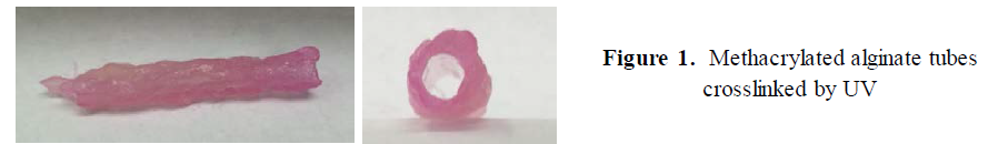
The alginate gel that was modified with methacrylic anhydride enabled the alginate gel to UV-crosslink. The modification also allowed the printed gel to maintain its structure while the seeded cells grow to form their own structure. UV-crosslinking is more desirable than the alternative ionic crosslinking method. UV-crosslinking creates a network of covalent bonds and can be crosslinked while printing. I was able to create a hollow tube using UV-crosslink methacrylated alginate as shown in Figure 1.
The optimal solution concentration for 3D printing was determined to be 7 % (w/v) in cell media. To sterilize the gels, I soaked the alginate for 30, 40 and 60 minutes in 70% ethanol, and I did not observe any contamination at 40 or 60 minutes. I concluded that 40 minutes was a sufficient time to sterilize the gel (size of 8 mm thick cylinder shape).
Discussion
In addition to crosslinking the methacrylated alginate, determining the right concentration for 3D printing and determining how to sterilize the gels, I learned that cell media may play a role in how well the methacrylated alginate gel crosslinks. For example, I tried dissolving the alginate in water instead of cell media, and the alginate did not crosslink as well in water. Specifically, the 3% methacrylated alginate crosslinked well with cell media, but not quite as well in water. The 7% gel crosslinked well enough to maintain its disk shape in the cell media, but crosslinked minimally in the water. The 5% gel did not crosslink in either. Furthermore, from these experiments, I learned that the 3% methacrylated alginate (the percentage meaning the ratio of methacrylated alginate to solution) crosslinks the best. In one of the experiments, I crosslinked the 3% gel for fifteen minutes, and it maintained its disk-shape for a number of days after being dissolved in water, and took a decent amount of pressure to break. However, the time under the UV light does make a difference because I also crosslinked a sample of 3% methacrylated alginate from the same batch under the UV light for only ten minutes, and it did not maintain its mechanical integrity as well because it broke into three pieces in five days or less. Even though the 3% methacrylated alginate crosslinked the best, it is not thick enough to print and maintain the shape necessary for a blood vessel. The 5% and 7% are thicker, so would print better, but take longer to crosslink. I think a possible way to help make the 5% and 7% methacrylated alginate crosslink better is to follow the pH curve more precisely by automating the syringe pump.
Conclusion
In conclusion, I learned that the crosslinking ability of the methacrylated alginate is affected by a number of factors, namely: the ratio of methacrylated alginate to solution, the type of solution, the time spent under the UV light, and how well the pH curve is followed.
References
1. Freeman, Inbar, Alon Kedem, and Smadar Cohen. “The Effect of Sulfation of Alginate Hydrogels on the Specific Binding and Controlled Release of Heparin-binding Proteins.” Biomaterials 29.22 (2008): 3260-268.
2. Jeon, Oju, Caitlin Powell, Shaoly M. Ahmed, and Eben Alsberg. “Biodegradable, Photocrosslinked Alginate Hydrogels with Independently Tailorable Physical Properties and Cell Adhesivity.” Tissue Engineering Part A 16.9 (2010): 2915-925.
3. Lee,KuenYong, and David J. Mooney. “Alginate: Properties andBiomedical Applications.”Progress in Polymer Science 37.1 (2012): 106-26.
4. Rouillard, Andrew D., Caroline M. Berglund, Jae Youn Lee,William J. Polacheck,Yvonne Tsui, Lawrence J. Bonassar, and Brian J. Kirby. “Methods for Photocrosslinking Alginate Hydrogel Scaffolds with High Cell Viability.” Tissue Engineering Part C: Methods 17.2 (2011): 173-79.